Disclaimer: This article is intended solely for informational and educational purposes only. It does not constitute medical advice.
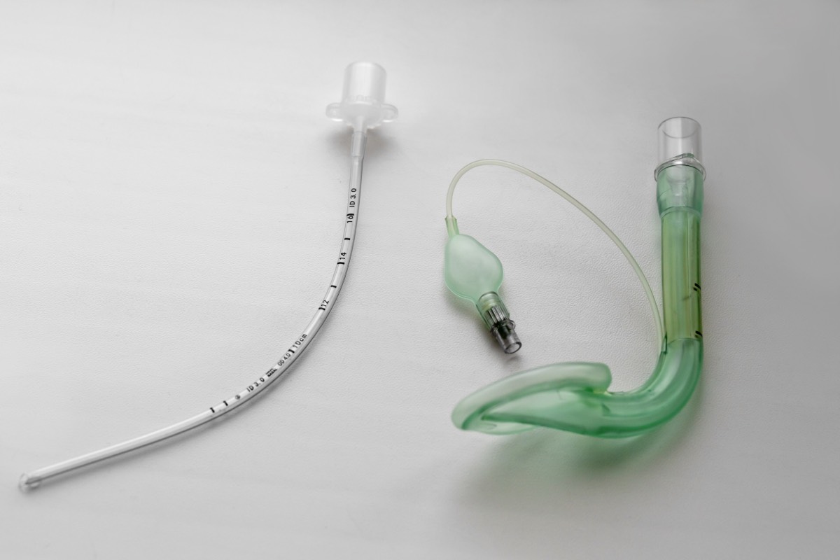
Deep extubation refers to the removal of the endotracheal tube from a patient who remains under a deep level of anesthesia and has not yet had their protective airway reflexes return. The technique is commonly employed to achieve a smoother emergence and minimize coughing, bucking, and hemodynamic stimulation associated with awake extubation. Although widely used in selected pediatric populations, deep extubation remains less common in adults because of concerns regarding airway obstruction, laryngospasm, aspiration, and loss of airway control. Literature suggests that when performed in carefully selected patients by experienced providers, deep extubation can be a safe and valuable anesthetic technique.
The primary advantage of deep extubation is the reduction of airway reflex activation during emergence. Coughing and bucking can increase intracranial, intraocular, and intrathoracic pressures, making deep extubation particularly attractive for ophthalmic, neurosurgical, head and neck, and certain plastic surgery procedures. Smooth emergence may also reduce sympathetic stimulation, resulting in less tachycardia and hypertension during recovery. Several studies have demonstrated decreased coughing and improved emergence characteristics compared with awake extubation.
Appropriate patient selection is the most important determinant of success. Ideal candidates have a low aspiration risk, an uncomplicated airway, and preserved spontaneous ventilation. Patients with known difficult airways, obesity, obstructive sleep apnea, gastroesophageal reflux disease, full stomachs, or significant pulmonary pathology may not be suitable candidates. Prospective studies in adult surgical patients have identified elevated body mass index and lower oxygen saturation before extubation as factors associated with an increased likelihood of respiratory complications following deep extubation.
Before extubation, the patient should be spontaneously ventilating with an adequate respiratory pattern and oxygen reserve. Neuromuscular blockade should be fully reversed, secretions thoroughly suctioned, and a clear plan for airway rescue established. Many practitioners maintain approximately 1 minimum alveolar concentration (MAC) of volatile anesthetic or an equivalent depth of intravenous anesthesia to ensure suppression of airway reflexes during extubation. Oral airways, jaw-thrust maneuvers, and supplemental oxygen are frequently used to support upper airway patency during the transition period. Continuous monitoring is essential until protective reflexes return and the patient demonstrates adequate airway control.
Potential complications include airway obstruction, breath holding, desaturation, laryngospasm, bronchospasm, and the need for reintubation. Airway obstruction is the most common adverse event and may result from loss of pharyngeal muscle tone during the period before emergence. Although these risks have limited the widespread adoption of deep extubation in adult anesthesia practice, available evidence suggests that complication rates remain relatively low when patients are carefully selected and extubation is performed by experienced providers. Most reported respiratory events are transient and respond to routine airway maneuvers such as jaw thrust, continuous positive airway pressure, or placement of an oral airway.
Deep extubation should be viewed as a specialized technique rather than a routine practice. Success depends on careful patient selection, meticulous preparation, adequate anesthetic depth, and the ability to rapidly manage airway complications if they occur. When these principles are followed, deep extubation can provide a smoother emergence and improve perioperative conditions for selected surgical procedures while maintaining an acceptable safety profile.
References
- Tsui BCH, Wagner A, Cave D, Elliott C, El-Hakim H, Malherbe S. Incidence of airway complications associated with deep extubation in adults. BMC Anesthesiol. 2020;20(1):299. DOI: 10.1186/s12871-020-01191-8
- Kim JY, Lee KH, Kim JY, et al. Deep vs. awake extubation and laryngeal mask airway removal in pediatric patients: a systematic review and meta-analysis. J Clin Med. 2018;7(10):353. DOI: 10.3390/jcm7100353
- Kaur M, Singh PM. Smooth extubation and smooth emergence techniques: a narrative review. J Anaesthesiol Clin Pharmacol. 2021;37(1):3-12. DOI: 10.1155/2021/8883257
- Cattano D, Rane M. Ventilation through an extraglottic tracheal tube: a technique for deep extubation and airway control. Br J Anaesth. 2017;118(6):959-960. DOI: 10.1093/bja/aex152