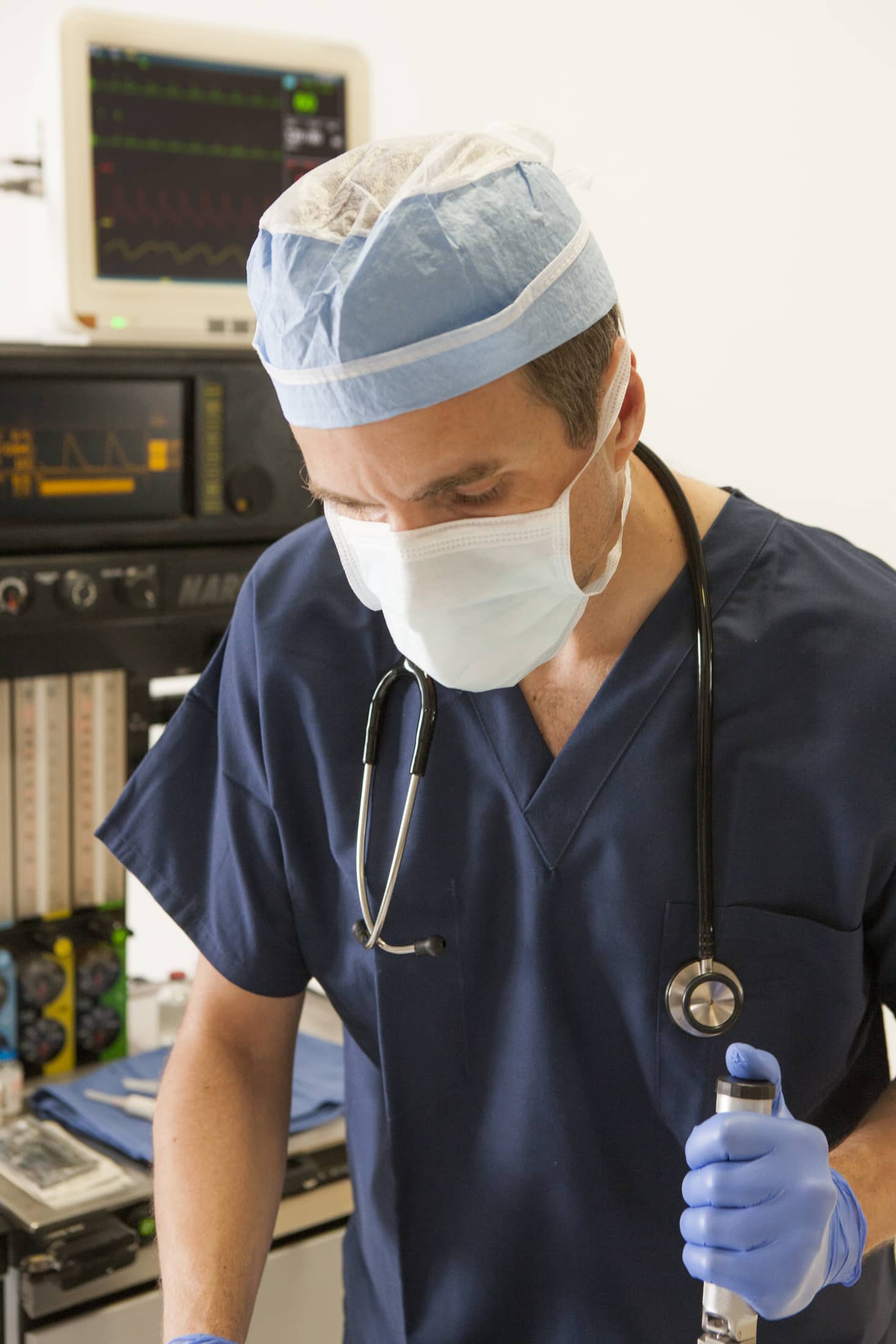
Like other out-of-OR locations, the magnetic resonance imaging (MRI) suite presents unique challenges to the anesthesiologist. Often remote from readily available backup both in terms of equipment and personnel common to the operating room suite, ensuring adequate foresight and preparation is essential to ensuring patient safety. Recent technological advances have facilitated the safe provision of anesthesia services in this setting.
While not all patients require anesthesia for MRI scans, their long duration and the enclosed system of the scanner make sedation helpful for children, developmentally delayed individuals, and claustrophobic or anxious adults to facilitate lying still. For some scans which require breath holds, i.e. for arterial mapping where even the slightest motion can interfere with image acquisition, general anesthesia may be necessary for those too young or unable to cooperate. Newer MRI scanners combine a wider bore with high-field systems to facilitate obese or claustrophobic patients’ comfort in the scanner, as well as decreasing the duration of the scan.
For those requiring some level of sedation for MRI scans, regimens range from anxiolytics such as midazolam, to propofol infusion based regimens, to general anesthesia with volatile gas. With deeper levels of sedation, monitoring airway patency is necessary. Given that the anesthesiologist must monitor the patient from outside the scan room, and the scanner often obscures direct view of the patient, direct visualization of chest rise or misting are ineffective measures. Use of end tidal carbon dioxide is a helpful tool and can be sampled from a nasal cannula or face mask, or from the ventilator circuit by way of mask, LMA or ETT. Often with the current technology this data, along with the patient’s vitals, can be transmitted to a remote monitor accessible to the anesthesiologist and even imported into an electronic medical record. Similarly, MRI-compatible infusion pumps have become advanced enough to be remotely controlled from outside the scan room, so that the level of sedation can be easily titrated.
Airway obstruction is of particular concern given the distance separating the anesthesiologist from the patient. Oral and nasal airways may be employed with deep enough sedation, and advanced airways with general anesthesia are again an option if the concern for airway compromise is high. One study described a prototype of the nasal vestibular airway (NVA) in anesthetized children undergoing MRI. The NVA is a nasal tube connected to an oxygen source and provides continuous positive pressure by means of a pressurized reservoir bag controlled by a valve and pressure gauge. The clinical advantage of this device over those already mentioned has not been formally studied.
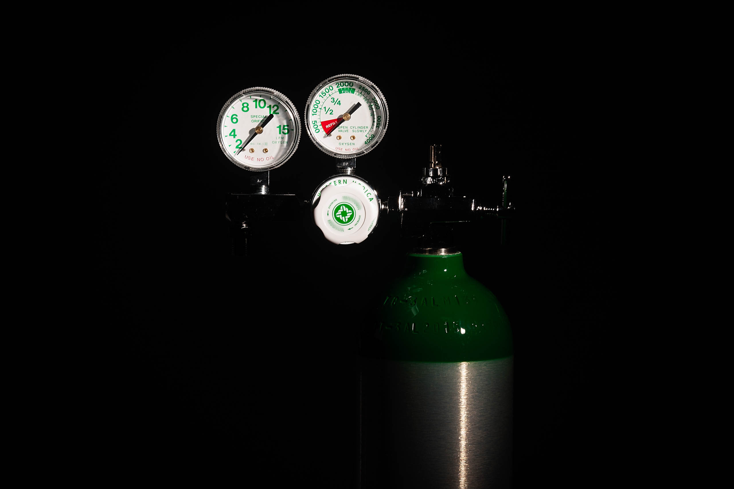
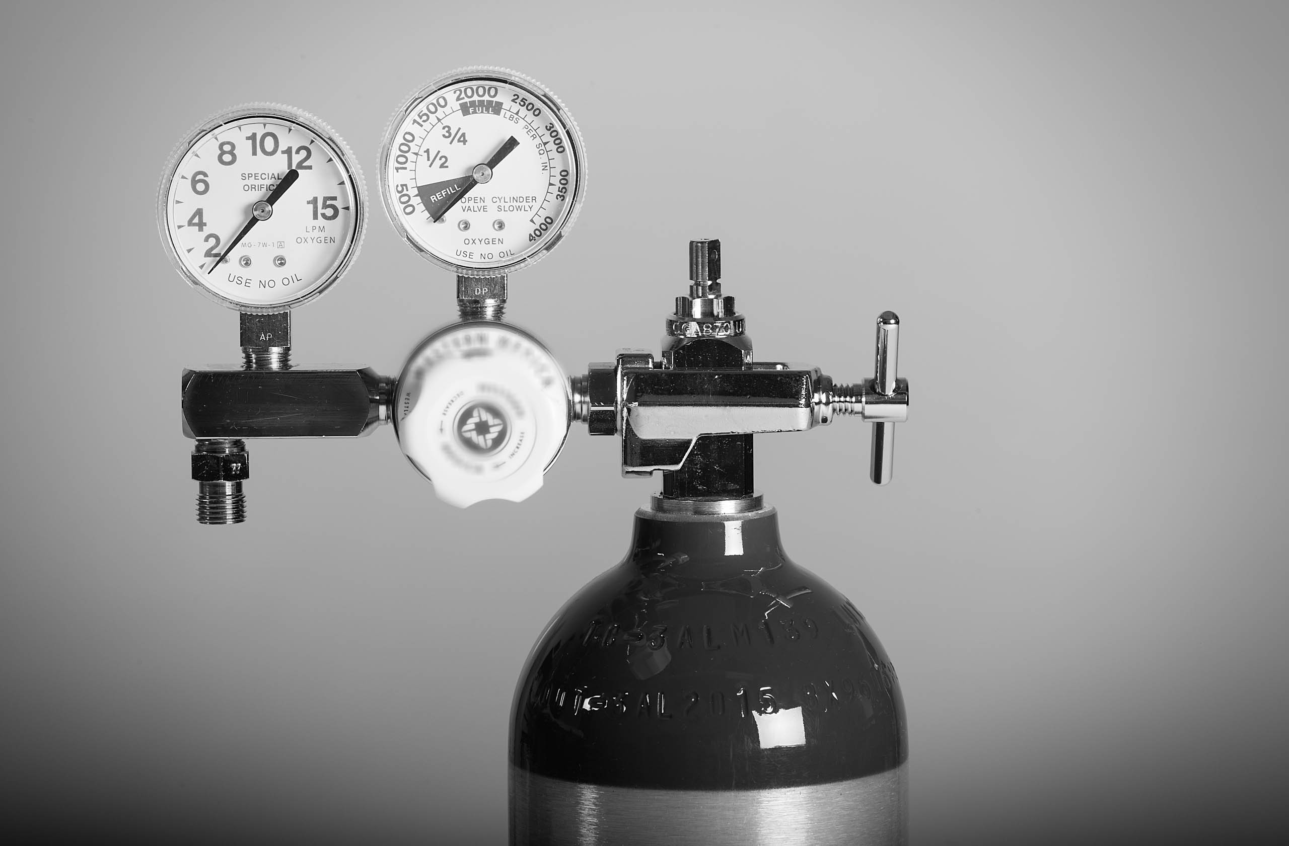
Compatibility of devices used in the scan room is of especial concern in MRI anesthesia. All monitors, infusion pumps, ventilators, gas tanks and other equipment should be clearly designated as MRI safe, with no ferromagnetic material (i.e. iron, nickel, cobalt) being allowed beyond the designated boundaries for fear of becoming dangerous projectiles. MRI compatible EKG electrodes and carbon fiber cables should be used to minimize the risk of burns, and coiling of wires should be avoided for the same reason. Similarly only MRI specific pulse oximeters should be used, as they contain heavy fiberoptic cables which do not overheat or coil onto themselves.
Anesthesia and sedation in the MRI suite presents a unique set of challenges to the anesthesiologist. Recent technological advances have improved safety and efficiency in providing patient care in this setting. Like other out-of-OR locations, adequate preparation and thoughtful consideration of environmental factors is of utmost importance.
References

Arthurs, Owen J; Sury, Michael. Anaesthesia or sedation for paediatric MRI: advantages and disadvantages. Current Opinion in Anaesthesiology. 26(4):489-494, August 2013.

Miller, Ronald D. (2015) Procedures Guided by Computed Tomography, Positron Emission Tomography, and Magnetic Resonance Imaging. In Miller’s Anesthesia (pp2659-2660). Philadelphia, PA: Elsevier.

Y Arlachov and R H Ganatra. Sedation/anaesthesia in paediatric radiology. The British Journal of Radiology 2012 85:1019, e1018-e1031


