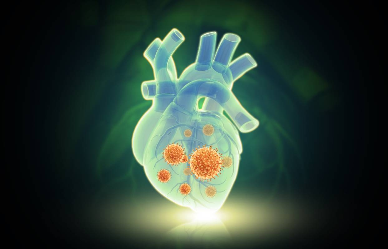
The largest meta-analysis of genetic susceptibility to COVID-19 infection included approximately 125,500 cases and over 2.5 million controls.

Genetic Susceptibility to COVID-19

By | Uncategorized | No Comments

Perhaps one of the most unique characteristics of the COVID-19 pandemic has been the large variability in infection-related morbidity and mortality. Disparities have been noted on several levels: between different ethnic groups and even within relatively homogenous demographics. Questions remain as to why some healthy, young adults have asymptomatic or mild infections while others require admission to the intensive care unit. For some researchers, these trends point towards the possibility of genetic and evolutionary determinants of COVID-19 susceptibility.1 

For patients with known genetic abnormalities, deletory effects on immune response to infection might be expected. For example, inborn errors of immunity, which include primary immunodeficiencies, increase susceptibility to a number of different infections, COVID-19 included, as well as autoimmune, inflammatory, and allergic diseases.2 A study performed by the COVID Human Genetic Consortium demonstrated that a myriad of monogenic predispositions which had been previously associated with influenza susceptibility also increased susceptibility to COVID-19. By performing both exomic and genomic sequencing on a large cohort of critically ill COVID-19 patients, the Consortium revealed thirteen such genes, including core immune genes TLR3, IRF7 and IRF9.2 

However, most individuals with genetic susceptibility to COVID-19 infection likely have a more complex etiology, orchestrated by multiple genes. The growing prevalence of genetic testing, both commercially and in healthcare settings, has made large-scale GWAS studies in the COVID-19 era possible. For example, the largest meta-analysis of genetic susceptibility to COVID-19 infection included approximately 125,500 cases and over 2.5 million controls.3 As a result of this mass study, the COVID-19 Host Genetics Initiative identified almost two dozen loci significantly associated with disease susceptibility or severity. One locus which was particularly prominent in their findings was on chromosome three, location 3p21.31 – a gene thought to encode an ACE-2 interacting sodium transporter.3 Given that SARS-CoV-2 targets the ACE-2 receptor on the cell membrane, the relationship between the 3p21.31 locus and increased disease susceptibility is consistent with known mechanisms of COVID-19 infection.  

As previously mentioned, significant discrepancies in the severity of SARS-CoV-2 infection have been noted across different ethnicities. Individuals of European ancestry tend to have reduced morbidity and mortality related to infection. While there is speculation that this disparity could be attributed to pervasive socioeconomic differences across racial groups, adjusting for these variables failed to fully ameliorate the difference for Black and South Asian populations, suggesting additional factors at play.4 Continued investigation revealed one unexpected predictor for susceptibility to COVID-19 infection: Neanderthal ancestry.5,6,7 A study conducted by the COVID-19 Host Genetics Initiative reported that a roughly 75kb haplotype, which is likely traceable to Neanderthal descent and is absent in African Homosapiens, is associated with a nearly 25 percent reduction in hospitalization risk due to COVID-19.6 The haplotype encodes antiviral enzymes which contribute to improved immunity. However, the beneficial effect of this haplotype may be negated by the inheritance of other, less advantageous genes: on chromosome 3, 3p21.31, a 50kb Neanderthal haplotype has been linked to increased severity of COVID-19 infection.7 This specific Neanderthal haplotype is at least three times more prevalent in South Asian populations and is associated with as much as a 60 percent increase in hospitalization.7 

Overall, much is still to be learned concerning evolutionary and genetic susceptibility to SARS-CoV-2 infection, particularly when it comes to the influence of Neanderthal ancestry on disease severity. However, continued investigation will continue to improve our understanding of the mechanisms underlying severe COVID-19 infection and may even help us to better identify at-risk populations or create more targeted curative treatments. 


References 


1 Kerner, G., & Quintana-Murci, L. (2022). The genetic and evolutionary determinants of COVID-19 susceptibility. European journal of human genetics : EJHG, 1–7. Advance online publication. https://doi.org/10.1038/s41431-022-01141-7 

2 Zhang, Q., Bastard, P., Liu, Z., Le Pen, J., Moncada-Velez, M., Chen, J., Ogishi, M., Sabli, I., Hodeib, S., Korol, C., Rosain, J., Bilguvar, K., Ye, J., Bolze, A., Bigio, B., Yang, R., Arias, A. A., Zhou, Q., Zhang, Y., Onodi, F., … Casanova, J. L. (2020). Inborn errors of type I IFN immunity in patients with life-threatening COVID-19. Science (New York, N.Y.), 370(6515), eabd4570. https://doi.org/10.1126/science.abd4570 

3 COVID-19 Host Genetics Initiative (2021). Mapping the human genetic architecture of COVID-19. Nature, 600(7889), 472–477. https://doi.org/10.1038/s41586-021-03767-x 

4 Williamson, E. J., Walker, A. J., Bhaskaran, K., Bacon, S., Bates, C., Morton, C. E., Curtis, H. J., Mehrkar, A., Evans, D., Inglesby, P., Cockburn, J., McDonald, H. I., MacKenna, B., Tomlinson, L., Douglas, I. J., Rentsch, C. T., Mathur, R., Wong, A., Grieve, R., Harrison, D., … Goldacre, B. (2020). Factors associated with COVID-19-related death using OpenSAFELY. Nature, 584(7821), 430–436. https://doi.org/10.1038/s41586-020-2521-4 

5 Zeberg, H., & Pääbo, S. (2020). The major genetic risk factor for severe COVID-19 is inherited from Neanderthals. Nature, 587(7835), 610–612. https://doi.org/10.1038/s41586-020-2818-3 

6 Pairo-Castineira, E., Clohisey, S., Klaric, L., Bretherick, A. D., Rawlik, K., Pasko, D., Walker, S., Parkinson, N., Fourman, M. H., Russell, C. D., Furniss, J., Richmond, A., Gountouna, E., Wrobel, N., Harrison, D., Wang, B., Wu, Y., Meynert, A., Griffiths, F., Oosthuyzen, W., … Baillie, J. K. (2021). Genetic mechanisms of critical illness in COVID-19. Nature, 591(7848), 92–98. https://doi.org/10.1038/s41586-020-03065-y 

7 Zeberg, H., & Pääbo, S. (2020). The major genetic risk factor for severe COVID-19 is inherited from Neanderthals. Nature, 587(7835), 610–612. https://doi.org/10.1038/s41586-020-2818-3 

Patient monitoring during anesthesia is critical to ensuring their well-being.

Blood Oxygen Monitoring During Anesthesia and Surgery

By | Uncategorized | No Comments

Patient monitoring during anesthesia is critical to ensuring their well-being. This includes monitoring a patient’s ventilation, circulation, body temperature, and oxygenation in both their inspired gas and in their blood. Blood oxygen monitoring during surgery is particularly important for maintaining adequate tissue and organ perfusion. This prevents the development of hypoxemia, hyperoxemia, and other such complications that can lead to negative outcomes 1,2. This is particularly problematic among patients who already have or are predisposed to respiratory problems, including but not limited to obstructive sleep apnea. To this end, blood oxygen monitoring methods such as pulse oximetry have been developed and successfully implemented during anesthesia. 


In order to gauge a patient’s oxygenation level, healthcare providers can assess (1) the saturation level of oxygen in hemoglobin via pulse oximetry, (2) the hematocrit, an indicator of hemoglobin concentration, and (3) the partial pressure of oxygen in arterial blood.  


Pulse oximetry noninvasively and continuously assesses the saturation of oxygen bound to hemoglobin. In particular, it measures the percentage of oxyhemoglobin and reduced hemoglobin present in arterial blood. Its downsides include the fact that it may be affected by many factors, including motion artifacts, ambient light, reduced peripheral blood flow (which can be caused by hypotension or vasoconstriction), electrical noise from surgical instruments, increased carboxyhemoglobin or methemoglobin concentrations in the blood, or darkly pigmented skin. 


Meanwhile, a patient’s hematocrit can be measured by a blood sample analysis usually prior to surgery, or, in some cases, from blood samples collected during surgery. In cardiac surgery in particular, it is critical to ensure that a patient’s hematocrit remains within a specified range 3. 


Finally, arterial blood gas measures probe the partial pressure of oxygen in arterial blood samples; this partial pressure of oxygen in arterial blood is directly linked to oxygen saturation levels in blood hemoglobin according to a linear relationship in most clinical contexts. 


Today, the standard protocol laid forth by the American Society of Anesthesiologists is to use a quantitative method of assessing oxygenation such as pulse oximetry 4. Adequate illumination and exposure of the patient are necessary to assess color, and an audible pulse tone and low threshold alarm should be used.  


A somewhat recent study sought to assess broadly conducted research on the effectiveness of pulse oximetry to identify hypoxemia and related events among a large general surgery population 5. The research team found no evidence that use of pulse oximetry affected the outcome of anesthesia. Specifically, routine continuous pulse oximetry monitoring neither reduced the rate of transfer to the intensive care unit nor decreased mortality. As such, the researchers concluded that the value of perioperative monitoring via pulse oximetry remains questionable as regards improved outcomes, effectiveness and efficiency.  


Intraoperative oxygen monitoring is critical to patient well-being, but further studies are required to improve our understanding of the clinical impact of intraoperative hypoxemia and the strategies that are most efficient in minimizing its occurrence.  


References 


  1. Ehrenfeld, J. M. et al. The incidence of hypoxemia during surgery: Evidence from two institutions. Can. J. Anesth. (2010). doi:10.1007/s12630-010-9366-5
  2. Karalapillai, D. et al. Frequency of hyperoxaemia during and after major surgery. Anaesth. Intensive Care (2020). doi:10.1177/0310057X20905320
  3. Kolotiniuk, N. V., Manecke, G. R., Pinsky, M. R. & Banks, D. Measures of Blood Hemoglobin and Hematocrit During Cardiac Surgery: Comparison of Three Point-of-Care Devices. J. Cardiothorac. Vasc. Anesth. (2018). doi:10.1053/j.jvca.2017.11.022
  4. American Society of Anestesiologist. Standards for Basic Anesthetic Monitoring. J. Chem. Inf. Model. (2020).
  5. Pedersen, T. et al. Pulse oximetry for perioperative monitoring. Cochrane Database of Systematic Reviews (2014). doi:10.1002/14651858.CD002013.pub3


The “No Surprises Act” attempts to remove patients from the center of these conflicts by establishing an independent dispute resolution process between insurers and providers.

Controversy Over “No Surprises Act” Implementation

By | Uncategorized | No Comments

Last December, Congress passed the “No Surprises Act” to shield patients from unexpected bills incurred when they receive medical treatment from providers outside their insurance networks. The bipartisan bill was applauded by groups like the American Hospitals Association,1 as well as consumers frustrated with the long history of “balance billing” in the United States — a process in which out-of-network providers bill individuals for the charges not covered by their insurance plans. The “No Surprises Act” attempts to remove patients from the center of these conflicts by establishing an independent dispute resolution process between insurers and providers.2 But as Congress finalizes the details of that process, many providers are protesting new guidelines they argue will benefit insurers at the expense of patients and providers.3 


Patients often encounter surprise bills in emergency settings, when they go to (or are taken to) a facility without consideration of their insurance plan due to a focus on quickly receiving definite care. A 2017 study found that patients who received surprise bills for emergency care paid ten times as much as those who did not, and that roughly 18 percent of emergency visits resulted in at least one out-of-network bill.4 But surprise bills can also appear when patients go to an in-network facility but receive care from an out-of-network provider, such as an anesthesiologist, surgical assistant, or ambulatory service.2 Roughly one in five privately insured patients undergoing an elective surgery at in-network hospitals received such a bill, according to a 2020 study. This was often attributed to anesthesiology expenses, with an average out-of-network bill of $1,219 in the study.4  


According to a new rule of the No Surprises Act, a particular benchmark — the qualifying payment amount (QPA) — should serve as a “starting point” in making payment determinations, as this number is “generally the plan or issuer’s median contracted rate for the same or similar service in the specific geographic area,” according to the Centers for Medicare and Medicaid Services (CMS).2 Parties can submit additional information if they wish to make an offer that deviates from the QPA-based offer, but it “must clearly demonstrate that the value of the item or service is materially different from the QPA,” according to the CMS.2  


Now, members of Congress are arguing that prioritizing the QPA over other considerations (physician’s training, quality of outcomes, local market share of the parties involved, etc.), will give insurers in upper hand in the dispute resolution process. Relying on the QPA may incentivize insurers to set artificially low payment rates, putting pressure on small practices, like anesthesiology practices, and limiting patients’ access to care. Over 150 lawmakers — nearly half of them Democrats, and some of them doctors themselves — signed a letter citing their opposition to the new rule.3 


Also speaking out against the rule are leading physician societies, including the American Society of Anesthesiologists (ASA). These societies argue that the QPA is calculated by the insurance companies “without meaningful oversight or transparency,” and therefore can be manipulated in their favor without reflecting actual payment rates, thereby undermining the spirit of the legislation, which emphasizes information sharing and the equal consideration of multiple factors.5 For this reason, some health care experts have emphasized that “rule makers should prioritize, strengthen, and highlight full historical context for arbiters and emphasize this information-sharing provision in final rulemaking for the arbitration process.”6 


Already, the ASA is protesting actions believed to be driven by insurers’ desires to maintain the upper hand in forthcoming disputes. According to the ASA, Blue Cross Blue Shield of North Carolina threatened in a letter to anesthesiology and physician practices that their contract and in-network status would be terminated unless they immediately agreed to payment reductions ranging from 10% to over 30%, with the No Surprises Act cited as driving the reduction. To many, this shows how the new rule may allow insurance companies to leverage their market power to prioritize their finances, pushing providers out of insurance networks or forcing them to accept lower rates along the way.7 It may particularly harm networks in rural and underserved areas by incentivizing insurers to push down the rates they pay to in-network providers.3 


Still, in some areas, a united group of providers may be stronger than the insurers in the market, allowing them to take the upper hand. The Congressional Budget Office also reports that patients may enjoy lower premiums, reduced by an estimated 1%, as a result of the act.3 If the act is passed with these provisions, it ultimately remains to be seen what effect it will have.


References 


  1. Detailed summary of No Surprises Act. (2021, January 14). American Hospitals Association. https://www.aha.org/advisory/2021-01-14-detailed-summary-no-surprises-act 
  1. Requirements Related to Surprise Billing; Part II interim final rule with comment period. (2021, September 30). Centers for Medicare and Medicaid Services. https://www.cms.gov/newsroom/fact-sheets/requirements-related-surprise-billing-part-ii-interim-final-rule-comment-period 
  1. McAuliff, M. (2021, November 17). Congressional doctors lead bipartisan revolt over policy on surprise medical bills. Kaiser Health News. https://khn.org/news/article/surprise-medical-bills-policy-congressional-doctors-backlash/ 
  1. Office of the Assistant Secretary for Planning and Evaluation. (2021, November 22). Evidence on Surprise Billing: Protecting Consumers with the No Surprises Act. U.S. Department of Health and Human Services. https://aspe.hhs.gov/sites/default/files/documents/acfa063998d25b3b4eb82ae159163575/no-surprises-act-brief.pdf 
  1. Nation’s frontline physicians denounce regulators’ implementation of key rule in No Surprises Act. (2021, October 1). American Society of Anesthesiologists. https://www.asahq.org/about-asa/newsroom/news-releases/2021/10/nations-frontline-physicians-denounce-regulators-implementation-of–key-rule-in-no-surprises-act 
  1. Koski-Vacirca, R., & Venkatesh, A. (2021, November 2). Rulemaking for health care affordability: Implementing the No Surprises Act. Health Affairs. https://www.healthaffairs.org/do/10.1377/hblog20211028.424036/full/  
  1. Lagasse, J. (2021, November 23). American Society of Anesthesiologists accuses BCBSNC of abusing No Surprises Act. Healthcare Finance News. https://www.healthcarefinancenews.com/news/american-society-anesthesiologists-accuses-bcbs-north-carolina-abusing-no-surprises-act  

Post-Extubation Airway Obstruction

By | Uncategorized | No Comments

Intubation is an acute respiratory intervention to ensure continued airflow through the upper airways and into the lungs. However, removal of the tube, also known as extubation, comes with an increased risk of airway obstruction. Partial obstruction, or stridor, can be supraglottic or glottic and is typically indicated by increased respiratory noise and struggle. Complete obstruction which can occur in the form of post-extubation airway obstruction typically points to a more extreme underlying condition and can be life-threatening. 

There are many risk factors which contribute to post-extubation airway obstruction. For example, one study performed by Tanaka et al. found that frequent endotracheal suctioning was correlated with increased incidence of stridor.1 Other risk factors include being female, prolonged intubation, and emergency intubation (as opposed to intubation in a controlled setting).2 These findings suggest that the structure of the laryngeal airway as well as the level of intubation-associated trauma contribute to the risk of post-extubation airway obstruction. 

Typically, conditions which involve a decrease in the airway lumen are associated with a higher risk of post-extubation airway obstruction. Laryngeal edema is commonly the underlying cause of partial or complete airway obstruction following extubation.3 In cases of extreme laryngeal edema, re-intubation may be necessary to re-establish respiratory flow. However, this is option is avoided if possible: re-intubation is often associated with a myriad of other complications, contributing to overall increased morbidity and mortality.4 Other types of lumen trauma, including ulcers and vocal cord damage, have also been found to be associated with post-extubation airway obstruction.3  

Although some instances of post-extubation airway obstruction are unpredictable, certain diagnostic tests can be performed prior to extubation to anticipate the integrity of the laryngeal airway. The gold standard for the past few decades has been the cuff leak test, which assesses the leak around the endotracheal tube when the cuff is deflated.5 The current threshold for a CLT is 110 mL of absolute volume. However, as authors Tokunaga et al. point out, there are a few issues with the current standard of CLT: for one, there is no way of validating the measurement, and no evaluation criteria have been established.6 Moreover, performing a CLT test increases the risk of patient-ventilator asynchrony.7 As an alternative, Tokunaga et. al report in their 2022 publication that measuring pressure above the cuff may serve as a less invasive alternative to the cuff link test to evaluate the risk of post-extubation airway obstruction.6 

The relative prevalence of post-extubation airway obstruction is not well known. Studies have estimated incidence rates of extubation-related stridor ranging from as low as 1.5 to as high as 26.3 percent.8 Similarly, studies have estimated the incidence of extubation-related laryngeal edema to be between five and 55 percent.5 This wide range points towards possible inconsistencies in the diagnosis and prevention of extubation-related stridor and laryngeal edema. 

Post-extubation airway obstruction is one of many complications associated with intubation. Although intubation is a commonly practiced intervention in emergency or hospital settings, it remains an invasive procedure associated with considerable discomfort. For that reason, risk assessments should be performed prior to any intubation, and then again prior to extubation. Tests such as the cuff leak test (or potentially the less invasive above-the-cuff pressure test) are critical to anticipating and avoiding life-threatening respiratory distress. 


References 


1 Tanaka, A., Uchiyama, A., Horiguchi, Y., Higeno, R., Sakaguchi, R., Koyama, Y., Ebishima, H., Yoshida, T., Matsumoto, A., Sakai, K., Hiramatsu, D., Iguchi, N., Ohta, N., & Fujino, Y. (2021). Predictors of post-extubation stridor in patients on mechanical ventilation: a prospective observational study. Scientific reports, 11(1), 19993. DOI: 10.1038/s41598-021-99501-8 

2 Shinohara, M., Iwashita, M., Abe, T., & Takeuchi, I. (2020). Risk factors associated with symptoms of post-extubation upper airway obstruction in the emergency setting. The Journal of international medical research, 48(5), 300060520926367. DOI: 10.1177/0300060520926367 

3 Colice, G. L., Stukel, T. A., & Dain, B. (1989). Laryngeal complications of prolonged intubation. Chest, 96(4), 877–884. DOI: 10.1378/chest.96.4.877 

4 Epstein, S. K., & Ciubotaru, R. L. (1998). Independent effects of etiology of failure and time to reintubation on outcome for patients failing extubation. American journal of respiratory and critical care medicine, 158(2), 489–493. DOI: 10.1164/ajrccm.158.2.9711045 

5 Zhou, T., Zhang, H. P., Chen, W. W., Xiong, Z. Y., Fan, T., Fu, J. J., Wang, L., & Wang, G. (2011). Cuff-leak test for predicting postextubation airway complications: a systematic review. Journal of evidence-based medicine, 4(4), 242–254. DOI: 10.1111/j.1756-5391.2011.01160.x 

6 Tokunaga, K., Ejima, T., Nakashima, T., Kuwahara, M., Narimatsu, N., Sagishima, K., Mizumoto, T., Sakagami, T., & Yamamoto, T. (2022). A novel technique for assessment of post-extubation airway obstruction can successfully replace the conventional cuff leak test: a pilot study. BMC anesthesiology, 22(1), 38. DOI: 10.1186/s12871-022-01576-x 

7 Sassoon C. (2011). Triggering of the ventilator in patient-ventilator interactions. Respiratory care, 56(1), 39–51. DOI: 10.4187/respcare.01006 

8 Pluijms, W. A., van Mook, W. N., Wittekamp, B. H., & Bergmans, D. C. (2015). Postextubation laryngeal edema and stridor resulting in respiratory failure in critically ill adult patients: updated review. Critical care (London, England), 19(1), 295. DOI: 10.1186/s13054-015-1018-2 

COVID-19 Rebound

By | Uncategorized | No Comments

In late May, CDC issued a health advisory regarding Paxlovid, an antiviral pill that is currently recommended for those with mild to moderate COVID-19 who are at high risk of progressing to severe disease. Preliminary data suggests that some patients who are treated with Paxlovid may experience a “rebound” or return of symptoms or test positivity a few days after initial recovery. The advisory outlines available information that is relevant for healthcare providers, public health agencies, and the general public. Importantly, reported cases of COVID-19 rebound thus far have been mild, and Paxlovid is currently still recommended for reducing the risk of hospitalization and death in at-risk individuals [1].


Paxlovid, also known as nirmatrelvir/ritonavir, is a prescription oral antiviral drug that reduces the risk of a COVID-19 case progressing to hospitalization and death in high-risk patients [1]. Populations considered to be at high risk include older adults, people with obesity, pregnant people, and those with certain medical conditions including diabetes, HIV, and cancer [2]. It has received emergency use authorization for people 12 and older. If eligible, the treatment consists of three pills taken two times per day for five days and should be started as soon as possible [1,2].


The advisory was made after a small number of case reports of COVID-19 rebound in patients with normal immune responses who completed the Paxlovid treatment course and seemed to fully recover – as measured by a negative test result [1,2]. Based on available data, experts do not believe that this phenomenon was due to reinfection with SARS-CoV-2 or to the virus developing resistance to the treatment, and other common respiratory illnesses were ruled out [1].


Cases so far have all been mild and resolved after a median of 3 days without needing additional treatment [1,2] However, CDC reported that there is a possibility that individuals experiencing COVID-19 rebound may be able to transmit the infection to others – additional research in this area is needed [1]. As a result, patients unfortunately need to restart their isolation period, following current guidelines [1,2].


Interestingly, in the Paxlovid clinical trial, a small number of participants had one or more positive test results after testing negative or an increase in the amount of SARS-CoV-2 detected by PCR after completing their treatment course, but this occurred in both the treatment group and the placebo group [1,2].


Case reports of potential rebound in COVID-19 patients generally (without any influence from Paxlovid) can be traced back to 2020. Researchers at the time raised the question of whether those cases represented reinfection or relapse. In one report, three older adults were diagnosed and hospitalized with COVID-19, clinically recovered and had a period without symptoms, and then returned to the hospital with confirmed infection several weeks after the first occurrence [3]. Another article reported on 11 patients who experienced a second episode days to weeks after the resolution of the first [4]. With additional data, it is now known that immunity due to vaccination and prior infection endures on the scale of months in people with normal immune responses, on average. However, abnormal immune responses to COVID-19 may allow a small percentage of people to be reinfected on a much shorter timescale. Whether COVID-19 itself is associated with potential rebound remains unclear.


Public health agencies and healthcare providers will no doubt be paying close attention to new cases of COVID-19 rebound moving forward and conducting additional research on the relative risks associated with Paxlovid and other treatments. For now, guidance on treatment and monitoring remains unchanged.


References


[1] “COVID-19 Rebound After Paxlovid Treatment.” CDC Health Alert Network, May 2022. Available online: https://emergency.cdc.gov/han/2022/pdf/CDC_HAN_467.pdf


[2] Bendix, A. “CDC warns of ‘Covid-19 rebound’ after taking Paxlovid antiviral pills.” NBC News, May 2022. Available online: https://www.nbcnews.com/health/health-news/cdc-warns-covid-19-rebound-taking-paxlovid-antiviral-pills-rcna30311


[3] Lafaie, L., et al. Recurrence or Relapse of COVID-19 in Older Patients: A Description of Three Cases. Journal of the American Geriatrics Society. 2020;68(10):2179-2183. doi: 10.1111/jgs.16728


[4]. Gousseff. M., et al. Clinical recurrences of COVID-19 symptoms after recovery: Viral relapse, reinfection or inflammatory rebound?. Journal of Infection. 2020;81(5):816-846. doi: 10.1016/j.jinf.2020.06.073