COVID-19 Rebound

By | Uncategorized | No Comments

In late May, CDC issued a health advisory regarding Paxlovid, an antiviral pill that is currently recommended for those with mild to moderate COVID-19 who are at high risk of progressing to severe disease. Preliminary data suggests that some patients who are treated with Paxlovid may experience a “rebound” or return of symptoms or test positivity a few days after initial recovery. The advisory outlines available information that is relevant for healthcare providers, public health agencies, and the general public. Importantly, reported cases of COVID-19 rebound thus far have been mild, and Paxlovid is currently still recommended for reducing the risk of hospitalization and death in at-risk individuals [1].


Paxlovid, also known as nirmatrelvir/ritonavir, is a prescription oral antiviral drug that reduces the risk of a COVID-19 case progressing to hospitalization and death in high-risk patients [1]. Populations considered to be at high risk include older adults, people with obesity, pregnant people, and those with certain medical conditions including diabetes, HIV, and cancer [2]. It has received emergency use authorization for people 12 and older. If eligible, the treatment consists of three pills taken two times per day for five days and should be started as soon as possible [1,2].


The advisory was made after a small number of case reports of COVID-19 rebound in patients with normal immune responses who completed the Paxlovid treatment course and seemed to fully recover – as measured by a negative test result [1,2]. Based on available data, experts do not believe that this phenomenon was due to reinfection with SARS-CoV-2 or to the virus developing resistance to the treatment, and other common respiratory illnesses were ruled out [1].


Cases so far have all been mild and resolved after a median of 3 days without needing additional treatment [1,2] However, CDC reported that there is a possibility that individuals experiencing COVID-19 rebound may be able to transmit the infection to others – additional research in this area is needed [1]. As a result, patients unfortunately need to restart their isolation period, following current guidelines [1,2].


Interestingly, in the Paxlovid clinical trial, a small number of participants had one or more positive test results after testing negative or an increase in the amount of SARS-CoV-2 detected by PCR after completing their treatment course, but this occurred in both the treatment group and the placebo group [1,2].


Case reports of potential rebound in COVID-19 patients generally (without any influence from Paxlovid) can be traced back to 2020. Researchers at the time raised the question of whether those cases represented reinfection or relapse. In one report, three older adults were diagnosed and hospitalized with COVID-19, clinically recovered and had a period without symptoms, and then returned to the hospital with confirmed infection several weeks after the first occurrence [3]. Another article reported on 11 patients who experienced a second episode days to weeks after the resolution of the first [4]. With additional data, it is now known that immunity due to vaccination and prior infection endures on the scale of months in people with normal immune responses, on average. However, abnormal immune responses to COVID-19 may allow a small percentage of people to be reinfected on a much shorter timescale. Whether COVID-19 itself is associated with potential rebound remains unclear.


Public health agencies and healthcare providers will no doubt be paying close attention to new cases of COVID-19 rebound moving forward and conducting additional research on the relative risks associated with Paxlovid and other treatments. For now, guidance on treatment and monitoring remains unchanged.


References


[1] “COVID-19 Rebound After Paxlovid Treatment.” CDC Health Alert Network, May 2022. Available online: https://emergency.cdc.gov/han/2022/pdf/CDC_HAN_467.pdf


[2] Bendix, A. “CDC warns of ‘Covid-19 rebound’ after taking Paxlovid antiviral pills.” NBC News, May 2022. Available online: https://www.nbcnews.com/health/health-news/cdc-warns-covid-19-rebound-taking-paxlovid-antiviral-pills-rcna30311


[3] Lafaie, L., et al. Recurrence or Relapse of COVID-19 in Older Patients: A Description of Three Cases. Journal of the American Geriatrics Society. 2020;68(10):2179-2183. doi: 10.1111/jgs.16728


[4]. Gousseff. M., et al. Clinical recurrences of COVID-19 symptoms after recovery: Viral relapse, reinfection or inflammatory rebound?. Journal of Infection. 2020;81(5):816-846. doi: 10.1016/j.jinf.2020.06.073

Investigation of Unusual Hepatitis Cases in Children

By | Uncategorized | No Comments

Public health officials in the US and internationally are actively investigating cases of severe, acute hepatitis without clear cause in children. Concerns were first escalated to WHO in early April based on reports of unusual cases across Scotland (1); as of mid-May, over 100 similar cases across roughly half of US states and territories have been identified, and over 200 probable cases have been identified globally (2). Though hepatitis is a disease that receives regular public health attention and research, this situation is notable for the severity and unknown etiology of cases, as well as the number of flagged cases (1, 2). 


Hepatitis is inflammation of the liver (3, 4). It can impair the liver’s normal functioning, which is to process nutrients, filter blood, remove toxins, and fight infections (3). Hepatitis is most commonly caused by viral infection, of which there are five known types (A through E), but it can also be caused by toxins, some medications, heavy alcohol use, and autoimmune conditions (3, 4). 


These cases warrant investigation due to their unusual nature. First, most affected children were previously healthy before falling ill. Second, cases drew the attention of specialists who then reported them to public health agencies due to their severity – around 90% of patients in the US required hospitalization, while around 15% required liver transplants, and 4% have died. Though hepatitis in general is not rare, this combination of healthy, young children falling so ill is extremely unusual (2). The liver is normally a relatively resilient organ – its regenerative ability is unique among solid organs, and liver failure is often a result of chronic conditions (5). Third, laboratory testing has excluded hepatitis virus as a cause, and so far, no etiology has been determined (1, 2). Reported symptoms include fatigue, loss of appetite, vomiting, diarrhea, abdominal pain, dark urine, light-colored stools, and jaundice (2). The working case definition used by WHO is someone presenting with “acute hepatitis (non-hepatitis viruses A, B, C, D, E) with aspartate transaminase (AST) or alanine transaminase (ALT) over 500 U/L, who is 10 years old and under, since 1 January 2022” (1). 


After the initial alert by UK authorities, public health officials began investigating health records to find similar cases that may not have been drawn significant attention on their own. Possible cases were found in Ireland and Spain soon after (1). Similarly in the US, the CDC was first notified of a cluster of cases in Alabama; subsequent investigation identified possible cases in other areas (2). 


Based on current data, the CDC is pursuing adenovirus as a leading possible cause. It is currently the only common factor that has been shared by public health agencies – many but not all of the identified children have tested positive for adenovirus (1, 2). Over 50 adenoviruses are known to be able to infect people, typically causing respiratory illness ranging from a cold to pneumonia and bronchitis, but sometimes causing inflammation in other organ systems (6). Genetic sequencing as a research tool for the cases with confirmed adenovirus infection has been limited by the fact that most samples did not have sufficient genetic material. The few cases that were partially or fully sequenced have all been adenovirus 41. However, this data point raises additional questions as adenovirus 41 has only been linked to hepatitis in immunocompromised children and never yet been linked to liver failure in otherwise healthy children (2). The majority of patients did not test positive for COVID-19 (1, 2). 


Ongoing research efforts seek to elucidate the etiology behind these unusual cases of severe hepatitis in children. Though the cases under investigation share key similarities, they have so far not been classified as an outbreak, and the epidemiological risk is thought to be low.  


References 


  1. World Health Organization (15 April 2022). Disease Outbreak News; Acute hepatitis of unknown aetiology – the United Kingdom of Great Britain and Northern Ireland. Available at: https://www.who.int/emergencies/disease-outbreak-news/item/acute-hepatitis-of-unknown-aetiology—the-united-kingdom-of-great-britain-and-northern-ireland  
  1. Brenda Goodman (6 May 2022). CDC investigating more than 100 cases of unexplained hepatitis in children, including 5 deaths. CNN. Available at: https://www.cnn.com/2022/05/06/health/hepatitis-kids-cdc-update/index.html 
  1. Centers for Disease Control and Prevention (28 July 2020). What is Viral Hepatitis?. Available at: https://www.cdc.gov/hepatitis/abc/index.htm 
  1. World Health Organization (n.d.). Hepatitis. Available at: https://www.who.int/health-topics/hepatitis 
  1. Michalopoulos, G.K., Bhushan, B. (2021). Liver regeneration: biological and pathological mechanisms and implications. Nat Rev Gastroenterol Hepatol, 18, 40–55. https://doi.org/10.1038/s41575-020-0342-4 
  1. Centers for Disease Control and Prevention (28 August 2019). Adenovirus. Available at: https://www.cdc.gov/adenovirus/index.html 


Automating Anesthesia Delivery

By | Uncategorized | No Comments

Each year, over 300 million patients worldwide undergo surgery, many of them requiring general anesthesia. Inhaled anesthetic agents, which are most common for general anesthesia, cost up to $1.2 billion annually [1]. Automating anesthesia delivery not only has the potential to reduce medical costs and minimize waste and greenhouse gas emissions, but may also make anesthesia delivery safer and more efficient.   


Research dating back to the 1990s has long investigated methods for the automated control of anesthetic delivery and ventilation during surgery. An anesthesia delivery system was developed as such to control fresh gas delivery, anesthetic delivery, and ventilation in order to regulate circuit volume, oxygen concentration, end-tidal anesthetic concentration, and end-tidal CO2 partial pressure according to an algorithm. This system capitalizes on the advantages of closed-circuit anesthesia without burdening anesthesiologists with complex control tasks. One study demonstrated that such a system was able to successfully maintain circuit volume, oxygen concentration, end-tidal anesthetic concentration, and end-tidal CO2 partial pressure in nearly all patients. However, a greater degree of variability was observed in certain measurements, including that of end-tidal anesthetic concentrations. 


More recently, research probed a novel automated anesthesia system for the closed-loop administration of intravenous anesthesia drugs for cardiac surgery which includes a cardiopulmonary bypass [2]. This anesthesia drug delivery system capitalizes on all three components of general anesthesia, including analgesia, hypnosis, and muscle relaxation. The trial enrolled twenty patients and found that robotic anesthesia was successful in 80%, with four cases experiencing a technical problem that required the anesthesiologist to take over manually for a short period. In the successful cases, the system demonstrated good clinical performance, suggesting that the completely automated closed-loop system tested could be safely and efficiently used for cardiac surgery requiring a cardiopulmonary bypass.  


Most recently, end-tidal control software has been so far offered by GE Healthcare, having received pre-market approval by the Food and Drug Administration (FDA) as a result of the United States-based, multi-center, multi-year MASTER-Anesthesia Trial which included over 200 patients [3]. This software semi-automates the delivery of anesthesia with the GE Aisys CS2 system, allowing anesthesia providers to directly set targets for end-tidal oxygen and anesthetic agent concentration. Once targets are set, the combined system swiftly reaches and maintains those targets, regardless of changes in the patient’s hemodynamic and metabolic patterns.  


Such software helps reduce greenhouse gas emissions and costs by cutting anesthetic agent waste – yielding a 44% drop in greenhouse gas emissions due to the more efficient use of anesthetic agents and a 27% drop in operating room costs according to a study in Australia, where the software was approved earlier [4]. Enabling anesthesia providers to set precise targets for oxygen and anesthetic agents, the software also increases workflow efficiencies by reducing manual keystrokes by up to 50%. 


Overall, the lack of specialized feedback sensors and the substantial degree of inter- and intra-individual variability in terms of responses to drug administration have limited the efficiency and reliability of closed-loop controllers in a clinical context. However, recent advances in sensing devices and nonlinear control theories have paved a promising path for automating anesthesia delivery. As such, technology in this field will likely continue to progress at a swift pace in the next few years – galvanizing the development of ever-more efficient forms of health care delivery. 


References 


  1. Weiser, T. G. et al. Size and distribution of the global volume of surgery in 2012. Bull. World Health Organ. (2016). doi:10.2471/blt.15.159293
  2. Zaouter, C. et al. The Feasibility of a Completely Automated Total IV Anesthesia Drug Delivery System for Cardiac Surgery. in Anesthesia and Analgesia (2016). doi:10.1213/ANE.0000000000001152
  3. FDA Approves Software to Semiautomate Anesthesia Delivery. Available at: https://www.medscape.com/viewarticle/971521.
  4. Tay, S., Weinberg, L., Peyton, P., Story, D. & Briedis, J. Financial and environmental costs of manual versus automated control of end-tidal gas concentrations. Anaesth. Intensive Care (2013). doi:10.1177/0310057×1304100116


COVID-19 Transmission Between Humans and Animals

By | Uncategorized | No Comments

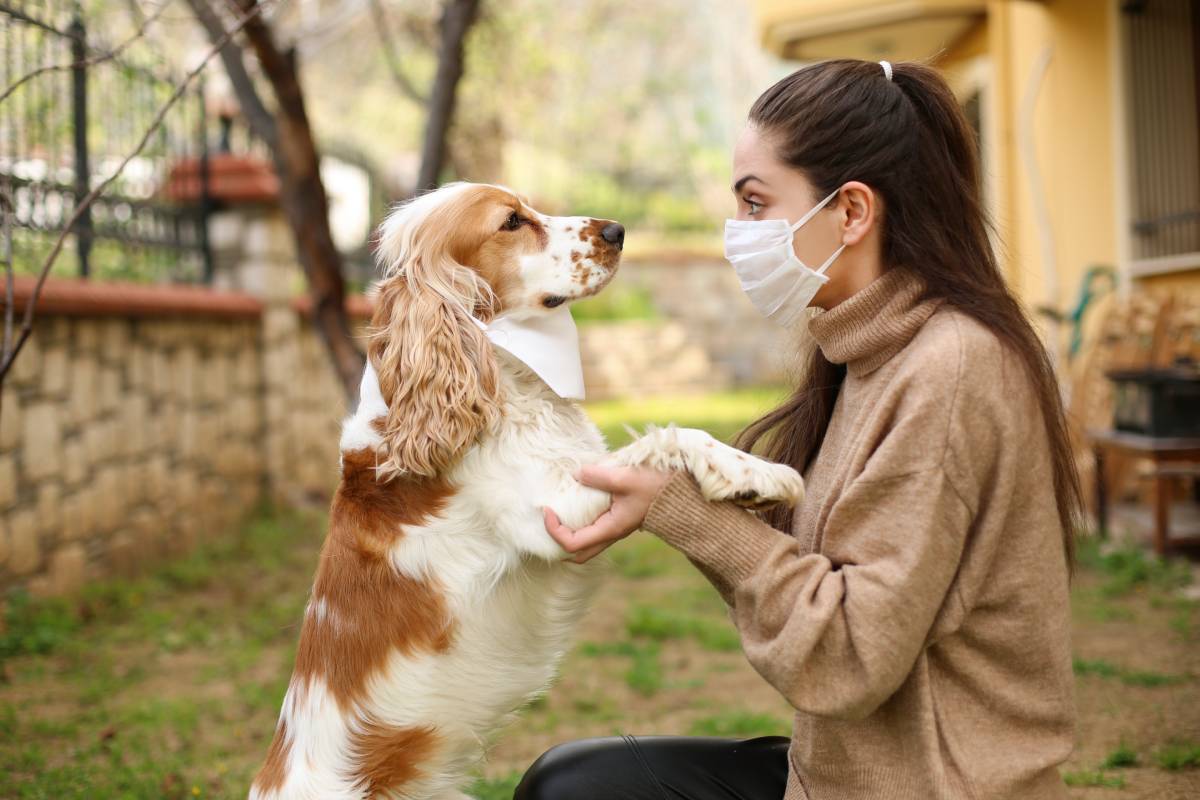
Coronaviruses are constantly evolving, switching and expanding host species, and, in the last few decades, novel coronaviruses have emerged in humans, domestic animals, and wildlife. The SARS-CoV-2 virus itself is thought by some to have originated in bats, crossing over to intermediate animal hosts (snake or pangolin) before infecting humans [1] and resulting in the global pandemic which to date has killed over 6 million individuals worldwide. While a definitive origin has not been found, one leading hypothesis of the genesis of COVID transmission into humans is a zoonotic origin, i.e. from animals. 

So far, SARS-CoV-2 has infected a number of animals. In addition to domestic animals, infections have been found in wild animals including minks, ferrets, big cats, great apes, and white-tailed deer[2]. A few of these species have been identified to date as transmitting SARS-CoV-2 to humans. In the first year of the pandemic, studies demonstrated that mink were capable of infecting humans with the SARS-CoV-2 virus [3,4]. Thereafter, pet hamsters were shown to transmit SARS-CoV-2 to humans [5]. In addition, a case has also recently been published of transmission to humans from white-tailed deer, who are particularly vulnerable to SARS-CoV-2 infection [6,7]. Conversely, COVID-19 can be transmitted from humans to animals. Critically, this is likely to amplify viral mutagenesis, and, in turn, result in the re-infection of humans with more virulent forms of the virus [8] 

High density environments that favor interspecies interactions – such as farms, markets, and animal shelters and kennels – have likely precipitated the emergence and transmission of coronaviruses by generating a large enough animal population susceptible to coronavirus circulation and spillover across species [9]. This is directly supported by findings that COVID-19 is far more prevalent in kennels than the rest of the dog population [10]. In addition, commercial agriculture has also led to many domestic animals living close to humans, possibly driving the emergence of key viruses from cattle (OC43) and camelids (229E and MERS). Finally, the rise of domestic animals populations has also similarly contributed the spike in COVID transmission from animals to humans. This trend is exacerbated by the fact that when animals are kept under poor stressful conditions (e.g. in overcrowded environments or frequently being transported), their experienced stress weakens their immune systems, rendering them even more susceptible to infections [11]  

Accordingly, the concept of One Health – a blanket strategy calling for “the collaborative efforts of multiple disciplines working locally, nationally, and globally, to attain optimal health for people, animals and our environment” – has been highlighted by COVID-19 and reports of human-animal transmission. To this end, first, personnel working closely with wildlife should be trained to implement measures that reduce the risk of disease transmission between and across people and animals, per World Health Organization (WHO) directives. This should include good hygiene practices for hunters and butchers [12]. Second, the public should be educated about what to do when entering in contact with wildlife. As a general precaution, people should not approach wild animals, instead choosing to notify wildlife authorities if an animal needs help. Third, it is critical to safely dispose of food and other human waste to avoid attracting wildlife, and, if possible, keeping domestic animals away from wildlife as well, all the while reducing people’s dependence on domestic animals. Finally, collaboration between veterinarians and wildlife authorities should be encouraged. Such work should include promoting the monitoring of wildlife and, in the context of COVID-19 specifically, the sampling of wild animals known to be potentially susceptible to SARS-CoV-2, sharing all genetic sequence data, and reporting confirmed animal SARS-CoV-2 cases, among others [2]. 

Efforts to monitor coronaviruses in the wild are currently underway (including the Viral Genome Project and PREDICT programs). These are key to identifying new viruses with zoonotic potential and dissecting potential spillover pathways [13]. Further research is warranted, in addition, to more fully understand and respond to cross-species transmission dynamics [14]. 


References  


  1. Mahdy, M. A. A., Younis, W. & Ewaida, Z. An Overview of SARS-CoV-2 and Animal Infection. Frontiers in Veterinary Science (2020). doi:10.3389/fvets.2020.596391
  2. Joint statement on the prioritization of monitoring SARS-CoV-2 infection in wildlife and preventing the formation of animal reservoirs. Available at: https://www.who.int/news/item/07-03-2022-joint-statement-on-the-prioritization-of-monitoring-sars-cov-2-infection-in-wildlife-and-preventing-the-formation-of-animal-reservoirs.
  3. Pomorska-Mól, M., Włodarek, J., Gogulski, M. & Rybska, M. Review: SARS-CoV-2 infection in farmed minks – an overview of current knowledge on occurrence, disease and epidemiology. Animal (2021). doi:10.1016/j.animal.2021.100272
  4. Munnink, B. B. O. et al. Transmission of SARS-CoV-2 on mink farms between humans and mink and back to humans. Science (80-. ). (2021). doi:10.1126/science.abe5901
  5. Haagmans, B. L. & Koopmans, M. P. G. Spreading of SARS-CoV-2 from hamsters to humans. Lancet 399, 1027–1028 (2022). doi:10.1038/s41586-021-04353-x
  6. Pickering, B. et al. Highly divergent white-tailed deer SARS-CoV-2 with potential deer-to-human transmission. bioRxiv 17 (2022). doi:10.1101/2022.02.22.481551
  7. Hale, V. L. et al. SARS-CoV-2 infection in free-ranging white-tailed deer. Nat. 2022 6027897 602, 481–486 (2021).
  8. He, S., Han, J. & Lichtfouse, E. Backward transmission of COVID-19 from humans to animals may propagate reinfections and induce vaccine failure. Environmental Chemistry Letters (2021). doi:10.1007/s10311-020-01140-4
  9. Plowright, R. K. et al. Pathways to zoonotic spillover. Nature Reviews Microbiology (2017). doi:10.1038/nrmicro.2017.45
  10. Naylor, M. J., Monckton, R. P., Lehrbach, P. R. & Deane, E. M. Canine coronavirus in Australian dogs. Aust. Vet. J. (2001). doi:10.1111/j.1751-0813.2001.tb10718.x
  11. Oreshkova, N. et al. SARS-CoV-2 infection in farmed minks, the Netherlands, April and May 2020. Eurosurveillance (2020). doi:10.2807/1560-7917.ES.2020.25.23.2001005
  12. CODE OF HYGIENIC PRACTICE FOR MEAT 1 CAC/RCP 58-2005. The National Institute for Communicable Diseases. Available at: https://www.nicd.ac.za/wp-content/uploads/2018/05/Code_of_Hygienic_Practice_for_Meat_CAC_RCP_58-2005.pdf
  13. Global Virome Project. Available at: https://www.globalviromeproject.org/.
  14. Animals and COVID-19 | CDC. Available at: https://www.cdc.gov/coronavirus/2019-ncov/daily-life-coping/animals.html.


Growth of Health Care Cost Fraction of GDP

By | Uncategorized | No Comments

Despite policymakers’ efforts to reduce health care costs via a number of different means since the 1980s, the growth of health care cost as a fraction of the United States GDP has continued a steady upward trend. Even after extracting out supplemental COVID-19 funding, the ratio of health care expenditure to gross domestic product (GDP) surpassed 18% in 2020. Despite high levels of spending at both the country and individual level, the U.S. trails behind many other high-income nations in key metrics of health and the functioning of the health care system. 


Since 1960, health care spending as a fraction of the GDP has risen by an average of 2.2 percentage points per decade – in sharp contrast to the average increase of 1.1 percentage points per decade in other high-income countries. Interestingly, this rise has been rather non-linear. Increases were higher than average in response to the establishment of Medicare and Medicaid in the 1960s, but lower than average in response to managed-care expansion in the 1990s and in the 2010s. The overall increase in health care costs as a fraction of the U.S. GDP can be linked to some long-term trends, however, including an aging population [3] and the lack of institutional budgets to limit and guide spending.  


Although health care spending has been funneled into hospital care, physician services, and other personal health care, life expectancy has not improved rapidly. On the contrary, increases in the health care cost fraction of the U.S. GDP have taken a toll on individual well-being. Most median-income American worker salary increases in the last few decades have been absorbed by increased taxes, health care premiums, and out-of-pocket expenses [4]. This is exacerbated by the fact that programs such as Medicare have no legislative limits on spending – this results in hospitals increasing prices in response to rising costs, meaning consumers have to pay higher insurance premiums. This system has precipitated patient bankruptcies, leading to unpaid bills and budget crises for state and even the federal governments.  


A number of interventions should be and have been made to reduce the cost of health care in the U.S. First, individual lifestyle factors can be improved to decrease the need to access health care. According to the American Medical Association (AMA), lifestyle modifications can reduce annual health care costs by nearly $2,700 per participant. Individuals and patients need to be well-educated about, and abide by, guidelines for a healthy lifestyle and available resources to this end, and access to and the effectiveness of interventions can be improved [5] 


At a systems level, reforming key administrative processes can also reduce costs. Prior authorization processing, managed care, deductibles and co-payments, standard electronic transactions, and bundled-payment programs are all areas that can be targeted. In many other Organization for Economic Cooperation and Development (OECD) countries, health care organizations adopt fixed budgets tied to tax revenue and general economic growth, resulting in a more stable ratio of health care expenditures to GDP [1] 


With the health care cost fraction of GDP reaching 18%, in stark contrast to the average 12% in other high-income countries, efforts need to continue to be made to reduce the growth of health care spending. Crucially however, this must be implemented in parallel with the adoption of novel, effective technologies and deliberate efforts to improve the quality of clinical care.  


References 

  1. Skinner, J., Cahan, E. & Fuchs, V. R. Stabilizing Health Care’s Share of the GDP. N. Engl. J. Med. 386, 709–711 (2022). doi:10.1056/NEJMp2114227
  2. Khan, T., Tsipas, S. & Wozniak, G. Medical Care Expenditures for Individuals with Prediabetes: The Potential Cost Savings in Reducing the Risk of Developing Diabetes. Popul. Health Manag. 20, 389–396 (2017). doi:10.1089/pop.2016.0134
  3. Why Are Americans Paying More for Healthcare? Available at: https://www.pgpf.org/blog/2022/02/why-are-americans-paying-more-for-healthcare.
  4. Auerbach, D. I. & Kellermann, A. L. A decade of health care cost growth has wiped out real income gains for an average US family. Health Aff. (2011). doi:10.1377/hlthaff.2011.0585
  5. The AMA can help you prevent type 2 diabetes | AMA Prevent Diabetes. Available at: https://amapreventdiabetes.org/.