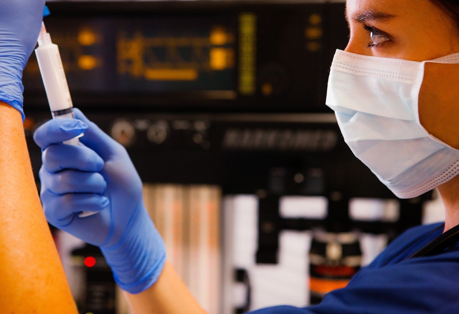
Anesthesia SimSTAT: Screen-Based Simulation Education

By | Health | No Comments

The practice of anesthesia, like its common comparison the aviation industry, has gained a great deal from simulation learning. Ideal for practicing high risk scenarios in a low risk setting, simulations have been shown in studies to accelerate skill acquisition, improve skill retention and reduce extinction of skills. Nontechnical skills such as task management, leadership, teamwork, situational awareness and decision making are also reinforced through simulation, and are particularly useful in emergency patient care. While additional data is needed to elucidate whether simulation makes a positive impact on patient outcomes, it has become integrated into residency and continuing medical education as a vital component of improving patient care and safety.

The evolution of the use of simulation in anesthesiology training has come a long way. Since the 1960s, life-like high-fidelity models have been in development. The incorporation of software based simulators using mathematical models of physiology and pharmacology, simulators were able to interact with mannequin models on a sophisticated level with lifelike responses. As advanced as these models are, they require upfront investment on the part of the simulation center and are limited in how many simulators can be accommodated at one time.

The ABA’s Maintenance of Certification in Anesthesiology Program (MOCA) initially required in Part 4 of their recredentialing process a certain number of hours to be spent in simulation at one of the ABA-endorsed centers nationwide, of which there are fewer than 50. The new MOCA 2.0 is a points based system allowing participants to chose among various activities, of which simulation is optional.

In the 2016 American Society of Anesthesiologists meeting, the ASA announced the upcoming release of a new screen-based simulation education program called Anesthesia SimSTAT. The program was developed in partnership with CAE Healthcare, a medical simulation company which has produced physical mannequin-based systems across a wide range of specialties. What is unique about SimSTAT is its ease of accessibility. It can be accessed via laptop via a web browser on both PC and Mac, requiring only an internet connection without need for plugins or installations.

SimSTAT will introduce on its release five learning modules at feature 3D graphics and interactive mechanics. A game based approach rewards points for successful navigation of the various cases. The user has unlimited attempts to improve competency in varied settings including the operating room, obstetric anesthesia, and the PACU. Rapid objective feedback is provided, along with practice guidelines and other resources.

Users are able to select an avatar to act as the anesthesiologist in five different scenarios. Three modes exist: orientation, which introduces the user to his environment; feedback, which allows the user to run through the scenario with frequent hints and guidance; and assessment, which runs the module without providing help or interruption. Nearly everything in the environment is interactive, with graphics for airway examination and sound bites for heart and lung sounds, fully functional ventilator dials, and an electronic medical record which updates when actions are taken by the anesthesiologist. The interface is intuitive, with the ability to access various actions either by clicking directly on the relevant object (e.g. clicking the IV pole to administer fluids or blood) or finding it on the navigation bar at the bottom of the screen.

Two scores are generated at the end of each module: an action score which represents the actions taken by the user and whether they were appropriate in timing and circumstance, and a physiological score which measures the deviations the patient’s vitals incur from acceptable parameters.

Due to release in 2017, SimSTAT promises many of the benefits of hands-on simulation combined with the convenience of being able to complete the modules anywhere, anytime. The program is currently in its beta testing phase and is preparing for general release in the upcoming months. Its target audience is primarily those seeking continuing medical education credits, specifically part 4 of MOCA; but there has been interest in using the modules as an educational platform for trainees as well. Those interested in receiving updates on its ongoing development can join the mailing list by contacting screenbasedsimulation@asahq.org.

spine surgery

Anesthesia Management of Spine Surgery

By | Health | No Comments

Patients undergo spine surgery for many different conditions, and these surgeries range from minimally invasive procedures to prolonged operations involving multiple spinal levels.  Anesthesia for spine surgery can present with many challenges, including difficult airway management, multiple patient cardiopulmonary comorbidities, significant blood loss, and pain control after surgery.  Anesthetic goals include providing optimal surgical conditions, reducing blood loss, avoiding spinal cord ischemia, and facilitating intraoperative neurophysiologic monitoring.

Preoperative evaluation includes special attention to airway anatomy and cervical spine instability.  Spinal deformities can result in compromised respiratory function, necessitating blood gas analysis and pulmonary function testing, and pulmonary hypertension could result from severe kyphoscoliosis.  Preoperative history and physical should include elicitation of sensorimotor deficits, which should be carefully documented.  Laboratory workup should include at least a baseline hemoglobin, coagulation factors, and type and screen.

Selection of anesthetic technique often is affected by neurophysiologic monitoring, which often includes motor and/or somatosensory evoked potentials that is facilitated with avoidance of volatile anesthetics and neuromuscular blockade.  A total intravenous anesthetic technique can be done with propofol and remifentanil infusions.  In anticipation of blood loss, two large bore intravenous catheters should be placed and connected to a fluid warmer. An arterial line might be required for continuous blood pressure monitoring and frequent blood draws. If vasoactive drugs are needed, a central venous catheter should be placed.  Awake intubation with a fiberoptic scope may be needed depending on anticipated airway challenges.  Induction and intubation are usually performed on the stretcher, and the patient is then repositioned prone onto the operating table with careful attention to position of the endotracheal tube.  Pressure points must be carefully padded and checked throughout the surgery to avoid optic injury and peripheral neuropathies, and jaw clenching necessitates a carefully secured bite block.

Many potential complications should be considered, especially during major spine surgeries.  The anesthesiologist should plan for the potential for massive blood loss, which may be reduced by the use of antifibrinolytic agents and controlled hypotension, depending on surgeon preference.  The anesthesiologist must carefully monitor and titrate blood pressures with consideration of the potential for spinal cord ischemia. Ischemic optic neuropathy is the most common cause of perioperative visual loss, a rare but devastating possibility.  Postoperative care may include admission to the intensive care unit, especially if the patient remains intubated due to concern for airway edema.

How Can Gene Therapy Impact You?

By | Health | No Comments

Gene therapy is an experimental technique for the treatment and prevention of disease by correcting its genetic cause through the introduction of a gene into targeted cells to either replace a mutated or missing gene, inactivate a defective gene, or introduce a new gene. The therapeutic benefits of gene editing as a medical intervention suggest new possibilities for the utilization of such technology for genetic enhancement, specifically increasing muscle strength, endurance, longevity, and intelligence. While there are currently no FDA-approved gene therapies in the U.S., a great amount of research is underway and the approval of therapies in or soon to enter clinical trials is expected in the near future.

A therapeutic gene enters its target cell by one of two types of gene transfer systems: viral vectors and non-viral carriers. Viral vectors are viruses rendered nonpathogenic and genetically-engineered to carry a therapeutic gene. DNA can also be transferred by non-viral carriers either by physical or chemical methods. Physical delivery systems use physical forces to weaken the target cell’s membrane and don’t involve a carrier. Chemical delivery systems introduce DNA into the nucleus through a carrier prepared by certain chemical reactions. Viral vectors’ high cost and ability to provoke an immune response led to the creation of these alternative delivery methods, however, viral vectors remain the most commonly used in gene therapy due to their efficiency. With gene transfer dependent on an efficient and safe gene delivery vehicle, developing such systems remains a challenge to the progress of gene therapy.

Since 1990, there have been over 1500 approved clinical gene therapy trials. Currently, gene therapy is only being tested on diseases with no known cures. Improvements from gene therapy treatments in diseases such as immunodeficiency diseases, cancer and eye disorders have been observed, however, effective and long-term treatment for certain anemia, hemophilia, cystic fibrosis, muscular dystrophy, and cardiovascular disease to name a few remain elusive.

Many candidates for genes that increase muscle strength have been studied for gene therapy to treat muscular dystrophies. Studies have indicated the potential for AAV-mediated follistatin gene therapy. Follistatin is an antagonist for myostatin, a negative regulator of muscle growth. Research has shown that blocking myostatin led to enhanced muscle strength and continuous muscle growth. Peroxisome proliferator-activated receptor-d (PPARd), another candidate for gene therapy, is also involved in muscle formation. Transgenic mice with an activated form of the gene demonstrated increased endurance and running capacity, and PPARd gene polymorphisms in humans are associated with cardiorespiratory fitness. The hormone erythropoietin (EPO) regulates erythrocyte production and is therapeutically used for the treatment of anemia.  EPO gene therapy delivers an extra EPO gene copy into the body, and the subsequent increase in erythrocyte levels augments endurance by increasing oxygen carrying capacity.

Instead of treating disease, genetic enhancement employs gene therapy strategies to improve human capacities. Studies on gene therapy to treat degenerative muscle disorders and certain anemia have elucidated possible techniques to enhance strength and endurance. The potential application of such discoveries calls into question the distinction between therapy and enhancement. This is especially apparent with gene therapy to increase “the time of life free of disease”, which potentiates life span extension. A study showed that telomerase reverse transcriptase (TERT) gene therapy in mice led to an increase in median lifespan, with telomerase acting “as a longevity gene in the context of the organism by preventing premature telomere attrition”. Although longevity has been found amenable to modulation in model systems, the nature and implications of both the epigenetic factors determining life span and the many genes whose frequencies vary with age remain unknown, hindering the feasibility of anti-aging gene therapy.

Genes that influence intelligence are also of great interest. Although many studies have been conducted, researchers have yet to conclusively identify any genes that dictate differences in intelligence among individuals. Studies suggest that 50% of this difference is attributed to genetic factors. Specific circuits within cortical areas have been hypothesized to encode learning and memory. Researchers have shown that the activation of protein kinase C (PKC) pathways in some of a circuit’s neurons activates the cortical circuit and thus improves learning: intracranial gene delivery of PKC into the postrhinal cortex of rats led to an improved learning rate.

The mode of gene editing techniques for both therapeutic and enhancement purposes poses ethical issues regarding the risks of such practices and the modification of the human genome. The goal of genetic enhancement additionally invokes concern, whereas the benefits of gene therapy to treat disease arguably prevail over the risk. The majority of the public approves of the clinical use of gene therapy in patients with serious diseases, and progress towards improving the efficacy and safety of gene therapy is underway. The possible applications of gene editing to improve patient health are promising, while nontherapeutic gene editing remains a theoretical possibility.

Ransomware

Rise of Ransomware in Healthcare

By | Health | No Comments

As more and more healthcare providers, anesthesiologists included, rely on online patient records and portable devices in their medical practices, the importance of understanding cyber-threats will only increase. Of particular interest is a malware called ransomware that has recently grown in popularity among cyber criminals. Its method of attack can appear alarmingly simple; once the victim opens an infected website or clicks on an infected advertisement, the malware can encrypt the files on the victim’s computer or connected digital network and render them unusable unless a ransom is paid for the data to be unencrypted. Ransomware can spread through the system at an alarming rate of less than three minutes and without the correct decryption key, the affected files may be permanently lost. While ransomware first appeared between 2005 and 2009, the recent advent of digital payment methods such as Bitcoin has allowed ransomware attacks to become more lucrative for criminals. While attacks on individuals used to focus on deception to trick victims into paying, attackers can now explicitly demand fees without fear of being tracked or identified.

Hospitals can be particularly vulnerable due to their dependence on daily access to patient records and lower focus on cybersecurity training. Publicized accounts of hospitals paying their ransoms may also contribute to their popularity as targets; in February of 2016, the Hollywood Presbyterian Medical Center in Los Angeles paid $17,000 in Bitcoin to have their access restored. A report by security company Solutionary for quarter 2 of 2016 found that 88% of ransomware detections were in the healthcare industry. Education, the second most affected industry, only accounted for 6% of detections.

Unfortunately, the number of attacks is only expected to increase as the ransomware industry continues to adapt and become harder to beat. The Los Angeles hospital was targeted using a ransomware variant called Locky, which employs phishing campaigns such as mass emails as well as network breaches to steal administrative credentials. Attacks using Locky also focus on locating the organization’s critical files; by inhibiting access to a shared server, they can bring a halt to the target organization’s activity.

In its report, Solutionary suggests that hospitals implement thorough backup and recovery processes as well as ensure that security software is up-to-date. Norton by Symantec, another security company, recommends that all users enable popup blockers and vet emails to prevent users from accidentally clicking on an infected source. In the event of ransomware infection, the company recommends victims avoid paying the criminals and also disconnect from the internet to prevent the transmission of personal information to the attackers. In the case of hospitals, the latter may entail shutting down network operations and reverting to paper records, as Medstar Health did in March 2016 in response to a suspected ransomware attack. If the ransomware variant is known, research can be done into whether there exist tools to bypass the encryption or at least mitigate the damage.

While there are methods of dealing with ransomware incidents, they can still take a toll on the hospital. Without a working patient portal, healthcare providers may find more of their time dedicated to admin tasks rather than patient care.  The best strategies may be those focused on preventative measures such as more rigorous cyber security training for employees. Examples of potential measures, adapted from those by suggested by Health Data Management, are as follows:

  • Educate the workforce on the risks of ransomware and potential sources.
  • Keep personal devices separate from the organization’s internal network to minimize security risk.
  • Create incident response protocols for cyberattacks and run drills to train workers on use of said protocols.

Hospitals should also ensure that their partners in hospital management are equally as dedicated to minimizing cybersecurity risks. Xenon Health has worked with multiple healthcare centers on enhancing their anesthesia services and understands the importance of privacy and cybersecurity to hospitals. Our overall goal is to improve the efficiency of anesthesia services as well as patient care and satisfaction and we fully embrace taking a multifaceted and technology-aware approach to achieve this goal.

Anesthetic Management in the Electrophysiology Lab

By | Health | No Comments

Cardiology procedures increasingly are being performed in the electrophysiology (EP) laboratory.  The rapid expansion of these diagnostic and therapeutic procedures and the increasing complexity and comorbidities these patients exhibit necessitate increasing involvement and expertise among anesthesia providers.  Providing anesthesia in a safe and effective manner in the EP laboratory requires encountering a variety of challenges ranging from patient factors to case complexity to resource availability.

Demand for anesthesia services in the cardiac EP lab has been growing over the past several years.  A survey in 2011 among cardiologists analyzed sedation practice in EP labs in the U.S. and showed that some patients transitioned into deep sedation or general anesthesia often without any supervision from an anesthesiologist due to lack of availability or difficulty in scheduling.  These procedures are becoming increasingly complex, and patients range from being very stable to being critically ill and undergo light sedation to general endotracheal anesthesia.  Therefore, anesthesia providers must be prepared to handle the full range of case complexity.

Procedures done in an EP laboratory include the following: placement of permanent pacemaker for symptomatic bradycardia or heart block, radiofrequency ablation for tachyarrhythmias, implantable cardiac defibrillator (ICD) placement for primary and secondary prevention of sudden cardiac death, and lead extractions for pacemaker or ICDs.  Anesthesiology services are warranted for patients who have the possibility of a difficult airway, obesity, obstructive sleep apnea, congestive heart failure, pulmonary disease, hemodynamic instability, neuromuscular disorders, or those who require a general anesthetic.

EP laboratories traditionally have been designed without any focus on the anesthesia provider.  Space and accessibility for the anesthesiologist can be rather limited, and usually the provider is physically located a significant distance away from the patient.  Equipment for imaging and monitors create physical and visual barriers, making it very difficult for the anesthesia provider to monitor vitals, assess the patient, and make adjustments as needed.  For example, once the procedure is under way, management of the airway and starting additional intravenous (IV) lines may be nearly impossible without significant disruption to the cardiologist.  Because of this, all lines including the airway circuit, IV access, and monitoring cables need to be carefully managed and secured prior to the procedure being started.  Additionally, further resources such as medications or anesthesia-related equipment must be anticipated and obtained well in advance due to the often-remote location of an EP laboratory.

To deal with these space constraints, the anesthesia provider must be well prepared.  All IV lines should have adequate length and often require adding tubing extensions.  The breathing circuit could be easily dislodged by rotating imaging equipment, so an extendable circuit should be used and carefully secured.  Monitoring cables and other special equipment such as arterial line transducers should be set up and remain in optimal position.  All of these factors must be considered early, and the anesthesiologist should be conservative in management decisions.  Additional invasive lines or monitors should be inserted in advance to prevent the need for rescue interventions during the procedure.

Most procedures in the EP laboratory are minimally invasive and do not require general anesthesia.  In order to come up with an anesthetic plan, the anesthesiologist must consider the procedure type and patient comorbidities as well as the preferences of the cardiologist and the patient.  To mitigate the pain of vascular access, which often is the most stimulating part of some EP procedures, liberal amounts of local anesthetic can be infiltrated.  Additionally, light sedation can be achieved by a propofol infusion supplemented by doses of opioids and/or benzodiazepines as needed. General anesthesia may be required for procedures such as ablation of atrial fibrillation, which can take several hours and requires the patient to be completely motionless.  The use of neuromuscular blocking agents should be discussed with the cardiologist in case phrenic nerve monitoring is needed.  Agents that can depress induction of tachyarrhythmias such as remifentanil or dexmedetomidine may be contraindicated in some EP procedures.  Before the procedure is begun, the anesthesia provider should have an understanding and awareness of the cardiologist’s plan for the case.

Complications in the EP laboratory include vascular access problems, oversedation, and airway difficulties.  Avoiding problems in the EP laboratory requires the anesthesiologist to understand the patient’s underlying condition, the procedure itself, and the potential complications that could arise.  Unfamiliarity with the equipment, remote location, and procedural and nursing staff can aggravate challenges to the anesthesia provider.  Close communication with the cardiologist and EP staff is critical for these cases.  Providing anesthesia services in the EP laboratory can present with many challenges, but with careful planning, excellent communication, and increasing familiarity with these complex procedures and patients, the anesthesiologist can provide safe and effective anesthesia care.