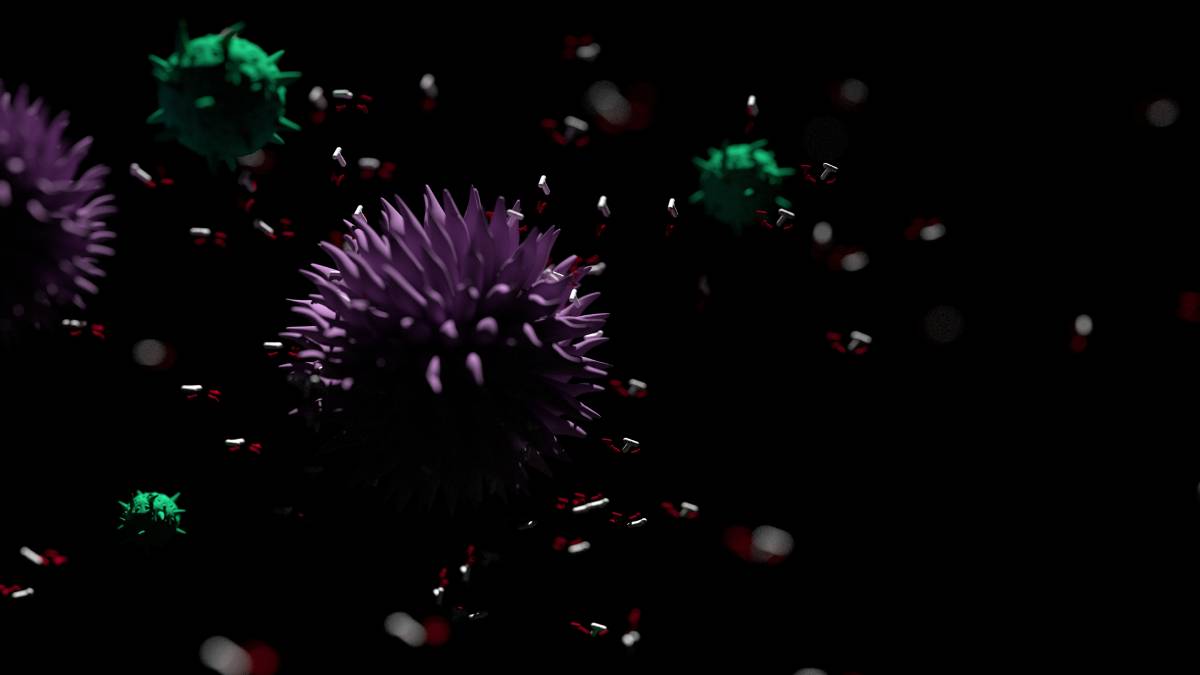
The human microbiome consists of all the microbes – bacteria, archaea, fungi, and even viruses – that live in and on human bodies and interact with human cells [1]. The term “microbiome” was first coined in 2001 [2] and it was discovered in subsequent years that this endogenous microbial population outnumbers human cells 10 to 1 and consists of 10,000 different microbial species [3]. This diversity helps account for the vast array of symbiotic human-microbial relationships scientists are only now beginning to uncover [4]. The absence of these beneficial microbes may heighten the risk for many diseases.
While microbial communities exist in the oral cavity, respiratory tract, and on the skin, microbes in the gastrointestinal tract have thus far been found to impact human health and physiology to the greatest extent [5]. Gut microbiota carry out important metabolic functions. For example, the digestion of xyloglucans, a type of cell wall polysaccharide found in lettuce and tomatoes, is performed by a mutant from the Bacteroides ovuatus species that inhabits the human gut [6]. Gut microbes also produce metabolites that signal endocrine cells to produce various hormones, some of which regulate insulin sensitivity, glucose tolerance, and fat storage [7].
Dysbiosis, the imbalance of natural microflora, can result from exposure to various environmental factors and has been implicated in several diseases [8]. Crohn’s disease and ulcerative colitis have been associated with an increase of Enterobacteriaceae and a loss of other symbiotic taxa [9]. Beyond gastrointestinal diseases, microbes responsible for the production of short-chain fatty acids such as butyrate can help lower the risk of heart disease [10]. Clostridium difficile infection, perhaps the most well-known example of dysbiosis, can occur when antibiotics wipe out colon bacteria that normally restrict C. difficile growth [11].
Characteristic of all these examples of dysbiosis is the reduction of microbial diversity. While it is difficult, perhaps impossible, to quantify the exact benefits generated by the various microbes that compose the microbiome, it is clear that an increase in diversity better prepares an organism to effectively respond to its environment. Reese and Dunn suggest implementing experiments whereby a population is “diluted” to produce individuals with reduced diversity, which can then be compared against normal individuals [12]. Mosca et al. implicate features of the Western lifestyle – eating behaviors, disruption of the circadian clock, and antibiotic consumption – in the loss of global microbial diversity and the resulting increase of allergies and inflammatory bowel diseases in the modern world. They explore reintroducing bacterial predators into the microbiota system to restabilize the microbial ecosystem [13].
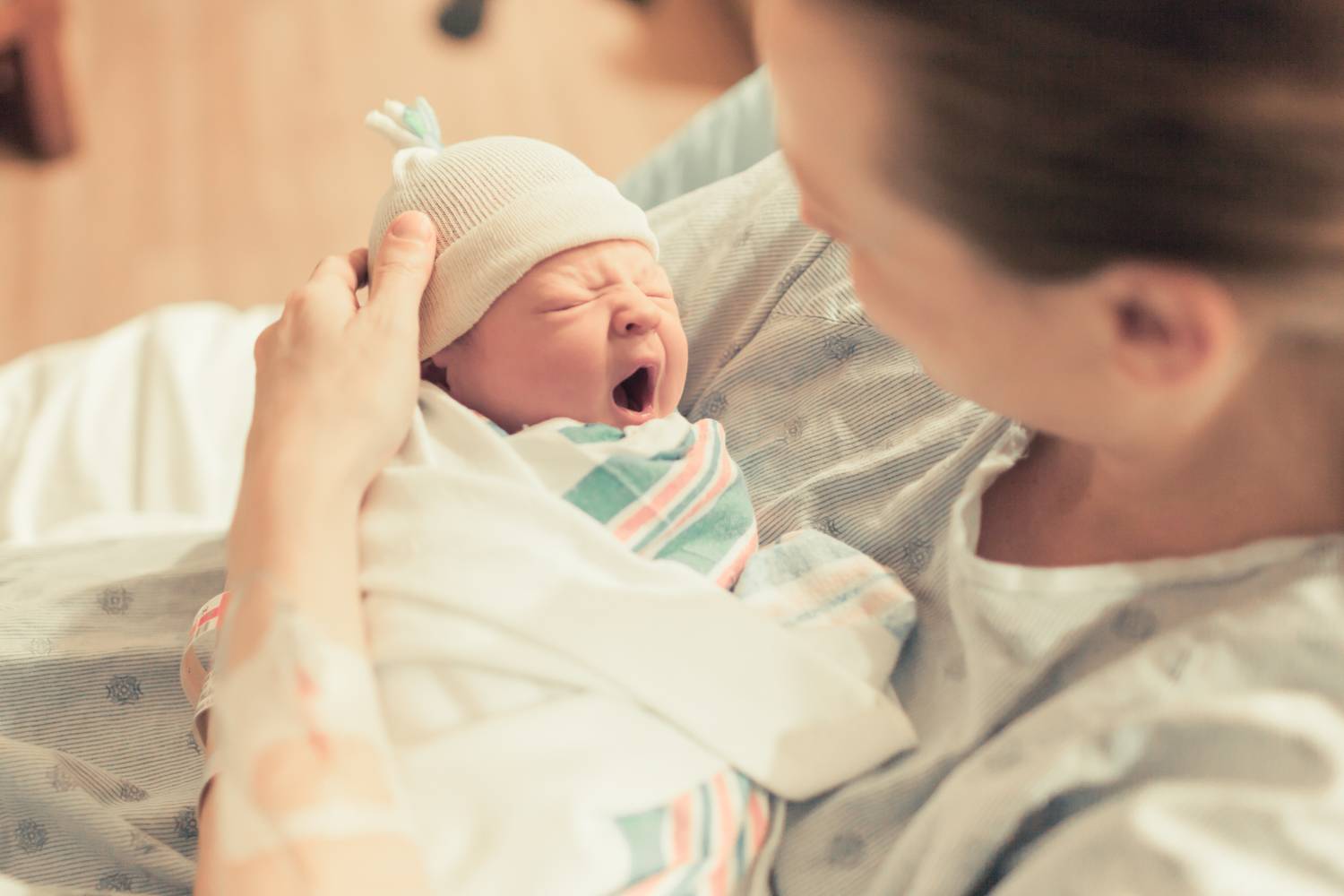
The effort to preserve and restore microbial diversity is far from simple. Bello et al. suggest several measures that can be implemented immediately – curtailing antibiotic use, limiting caesarean sections, promoting breastfeeding – but they acknowledge that ultimately, a more ambitious strategy to systematically identify and reintroduce symbiotic bacteria is needed [14]. Part of the problem stems from the difficulty of sampling and identifying the thousands of microbes inhabiting humans from different populations. A recently developed technology called Cell Alive System uses magnetic fields and mechanical vibrations to uniformly cool microbial samples, which gives them the best chance of survival in vitro. The decrease in microbial diversity is an urgent public health problem that demands our attention. As Dr. Martin Blaser, chair of the Human Microbiome at Rutgers University puts it, “The first point is to stop the damage, then rebuild” [15].
References
- “The Microbiome.” The Nutrition Source, 1 May 2020, www.hsph.harvard.edu/nutritionsource/microbiome/.
- Prescott, Susan L. “History of Medicine: Origin of the Term Microbiome and Why It Matters.” Human Microbiome Journal, vol. 4, 2017, pp. 24–25., doi:10.1016/j.humic.2017.05.004.
- “NIH Human Microbiome Project Defines Normal Bacterial Makeup of the Body.” National Institutes of Health, U.S. Department of Health and Human Services, 31 Aug. 2015, www.nih.gov/news-events/news-releases/nih-human-microbiome-project-defines-normal-bacterial-makeup-body.
- Ogunrinola, Grace A., et al. “The Human Microbiome and Its Impacts on Health.” International Journal of Microbiology, vol. 2020, 2020, pp. 1–7., doi:10.1155/2020/8045646.
- Barton, Wiley, et al. “Metabolic Phenotyping of the Human Microbiome.” F1000Research, vol. 8, 2019, p. 1956., doi:10.12688/f1000research.19481.1.
- Larsbrink, Johan, et al. “A Discrete Genetic Locus Confers Xyloglucan Metabolism in Select Human Gut Bacteroidetes.” Nature, vol. 506, no. 7489, 2014, pp. 498–502., doi:10.1038/nature12907.
- Martin, Alyce M., et al. “The Influence of the Gut Microbiome on Host Metabolism Through the Regulation of Gut Hormone Release.” Frontiers in Physiology, vol. 10, 2019, doi:10.3389/fphys.2019.00428.
- Carding, Simon, et al. “Dysbiosis of the Gut Microbiota in Disease.” Microbial Ecology in Health & Disease, vol. 26, 2015, doi:10.3402/mehd.v26.26191.
- Durack, Juliana, and Susan V. Lynch. “The Gut Microbiome: Relationships with Disease and Opportunities for Therapy.” Journal of Experimental Medicine, vol. 216, no. 1, 2018, pp. 20–40., doi:10.1084/jem.20180448.
- Baxter, Nielson T., et al. “Dynamics of Human Gut Microbiota and Short-Chain Fatty Acids in Response to Dietary Interventions with Three Fermentable Fibers.” MBio, vol. 10, no. 1, 2019, doi:10.1128/mbio.02566-18.
- “C. Difficile Infection.” American College of Gastroenterology, 6 Feb. 2020, gi.org/topics/c-difficile-infection/.
- Reese, Aspen T., and Robert R. Dunn. “Drivers of Microbiome Biodiversity: A Review of General Rules, Feces, and Ignorance.” MBio, vol. 9, no. 4, 2018, doi:10.1128/mbio.01294-18.
- Mosca, Alexis, et al. “Gut Microbiota Diversity and Human Diseases: Should We Reintroduce Key Predators in Our Ecosystem?” Frontiers in Microbiology, vol. 7, 2016, doi:10.3389/fmicb.2016.00455.
- Bello, Maria G. Dominguez, et al. “Preserving Microbial Diversity.” Science, vol. 362, no. 6410, 2018, pp. 33–34., doi:10.1126/science.aau8816.
- “Disappearance of the Human Microbiota: How We May Be Losing Our Oldest Allies.” ASM.org, asm.org/Articles/2019/November/Disappearance-of-the-Gut-Microbiota-How-We-May-Be.