
Cardiology procedures increasingly are being performed in the electrophysiology (EP) laboratory. The rapid expansion of these diagnostic and therapeutic procedures and the increasing complexity and comorbidities these patients exhibit necessitate increasing involvement and expertise among anesthesia providers. Providing anesthesia in a safe and effective manner in the EP laboratory requires encountering a variety of challenges ranging from patient factors to case complexity to resource availability.
Demand for anesthesia services in the cardiac EP lab has been growing over the past several years. A survey in 2011 among cardiologists analyzed sedation practice in EP labs in the U.S. and showed that some patients transitioned into deep sedation or general anesthesia often without any supervision from an anesthesiologist due to lack of availability or difficulty in scheduling. These procedures are becoming increasingly complex, and patients range from being very stable to being critically ill and undergo light sedation to general endotracheal anesthesia. Therefore, anesthesia providers must be prepared to handle the full range of case complexity.
Procedures done in an EP laboratory include the following: placement of permanent pacemaker for symptomatic bradycardia or heart block, radiofrequency ablation for tachyarrhythmias, implantable cardiac defibrillator (ICD) placement for primary and secondary prevention of sudden cardiac death, and lead extractions for pacemaker or ICDs. Anesthesiology services are warranted for patients who have the possibility of a difficult airway, obesity, obstructive sleep apnea, congestive heart failure, pulmonary disease, hemodynamic instability, neuromuscular disorders, or those who require a general anesthetic.
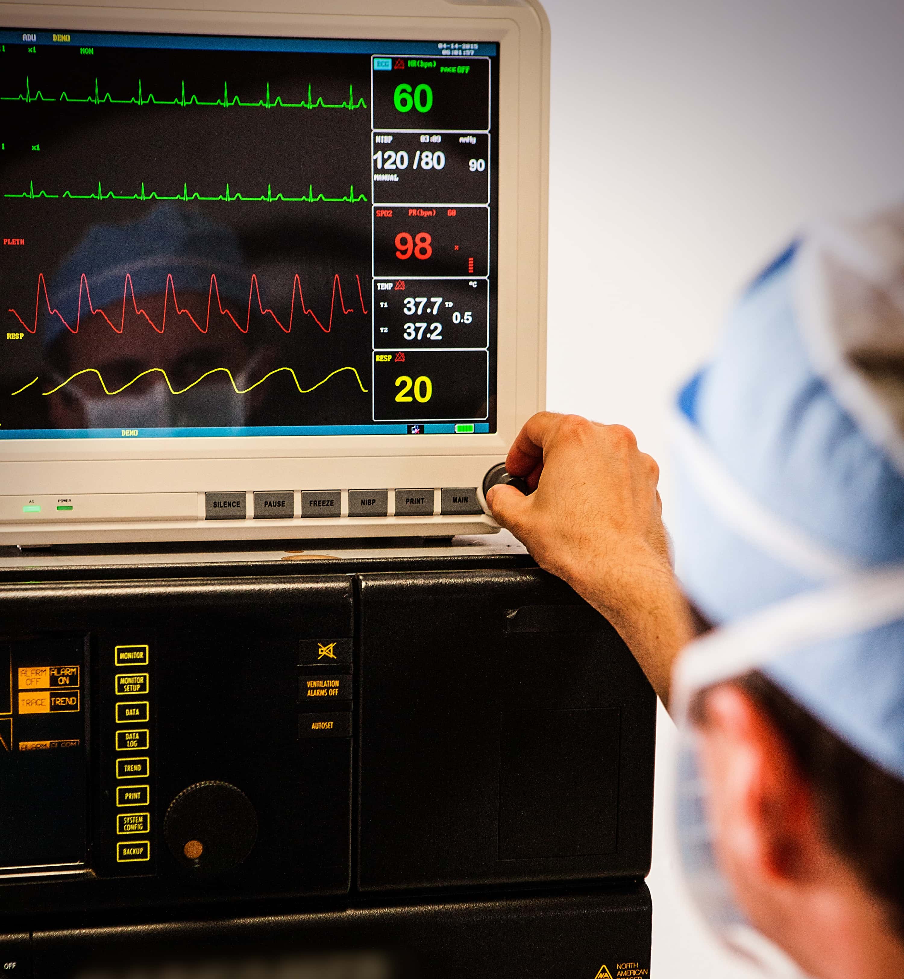
EP laboratories traditionally have been designed without any focus on the anesthesia provider. Space and accessibility for the anesthesiologist can be rather limited, and usually the provider is physically located a significant distance away from the patient. Equipment for imaging and monitors create physical and visual barriers, making it very difficult for the anesthesia provider to monitor vitals, assess the patient, and make adjustments as needed. For example, once the procedure is under way, management of the airway and starting additional intravenous (IV) lines may be nearly impossible without significant disruption to the cardiologist. Because of this, all lines including the airway circuit, IV access, and monitoring cables need to be carefully managed and secured prior to the procedure being started. Additionally, further resources such as medications or anesthesia-related equipment must be anticipated and obtained well in advance due to the often-remote location of an EP laboratory.
To deal with these space constraints, the anesthesia provider must be well prepared. All IV lines should have adequate length and often require adding tubing extensions. The breathing circuit could be easily dislodged by rotating imaging equipment, so an extendable circuit should be used and carefully secured. Monitoring cables and other special equipment such as arterial line transducers should be set up and remain in optimal position. All of these factors must be considered early, and the anesthesiologist should be conservative in management decisions. Additional invasive lines or monitors should be inserted in advance to prevent the need for rescue interventions during the procedure.
Most procedures in the EP laboratory are minimally invasive and do not require general anesthesia. In order to come up with an anesthetic plan, the anesthesiologist must consider the procedure type and patient comorbidities as well as the preferences of the cardiologist and the patient. To mitigate the pain of vascular access, which often is the most stimulating part of some EP procedures, liberal amounts of local anesthetic can be infiltrated. Additionally, light sedation can be achieved by a propofol infusion supplemented by doses of opioids and/or benzodiazepines as needed. General anesthesia may be required for procedures such as ablation of atrial fibrillation, which can take several hours and requires the patient to be completely motionless. The use of neuromuscular blocking agents should be discussed with the cardiologist in case phrenic nerve monitoring is needed. Agents that can depress induction of tachyarrhythmias such as remifentanil or dexmedetomidine may be contraindicated in some EP procedures. Before the procedure is begun, the anesthesia provider should have an understanding and awareness of the cardiologist’s plan for the case.
Complications in the EP laboratory include vascular access problems, oversedation, and airway difficulties. Avoiding problems in the EP laboratory requires the anesthesiologist to understand the patient’s underlying condition, the procedure itself, and the potential complications that could arise. Unfamiliarity with the equipment, remote location, and procedural and nursing staff can aggravate challenges to the anesthesia provider. Close communication with the cardiologist and EP staff is critical for these cases. Providing anesthesia services in the EP laboratory can present with many challenges, but with careful planning, excellent communication, and increasing familiarity with these complex procedures and patients, the anesthesiologist can provide safe and effective anesthesia care.